Microbiology Case Study: A Novel Anaerobic Pathogen Causing Septic Arthritis

Clinical history

A 65 year old man with diabetes mellitus type 2 presented to the emergency department (ED) for left hip pain. He has a remote history of avascular necrosis of bilateral hips of unknown etiology for which he received a bilateral total hip arthroplasty and subsequent multiple revisions due to hardware failure several years ago. He initially presented to an urgent care clinic a few months prior for “noise with movement” of the left hip and mild lower back pain. Plain radiographs of the left hip in comparison to his prior imaging were unremarkable and he was subsequently discharged. Repeat imaging at a follow-up visit at the orthopedic clinic showed mild superior migration of the femoral head bilaterally secondary to periprosthetic osteolysis of the joint headliner. He was scheduled for surgery however presented to the ED prior to his scheduled appointment with severe crushing left hip pain and restricted joint mobilization. He denied fevers, chills, night sweats, or any other recent infections. The left hip was aspirated yielding 10cc of dark black fluid and a stat gram stain was ordered.

Laboratory identification

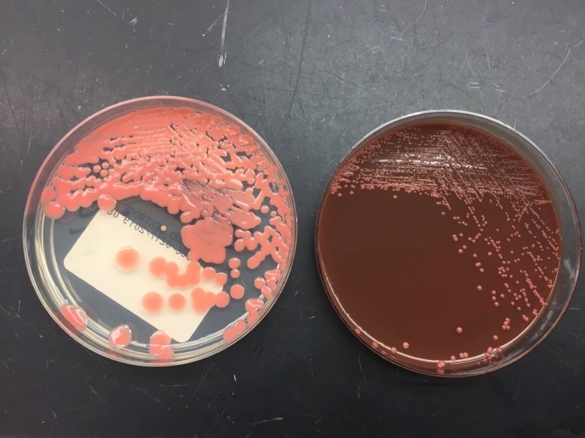
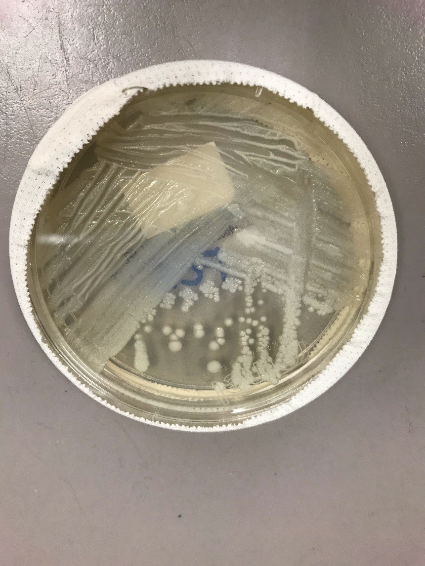
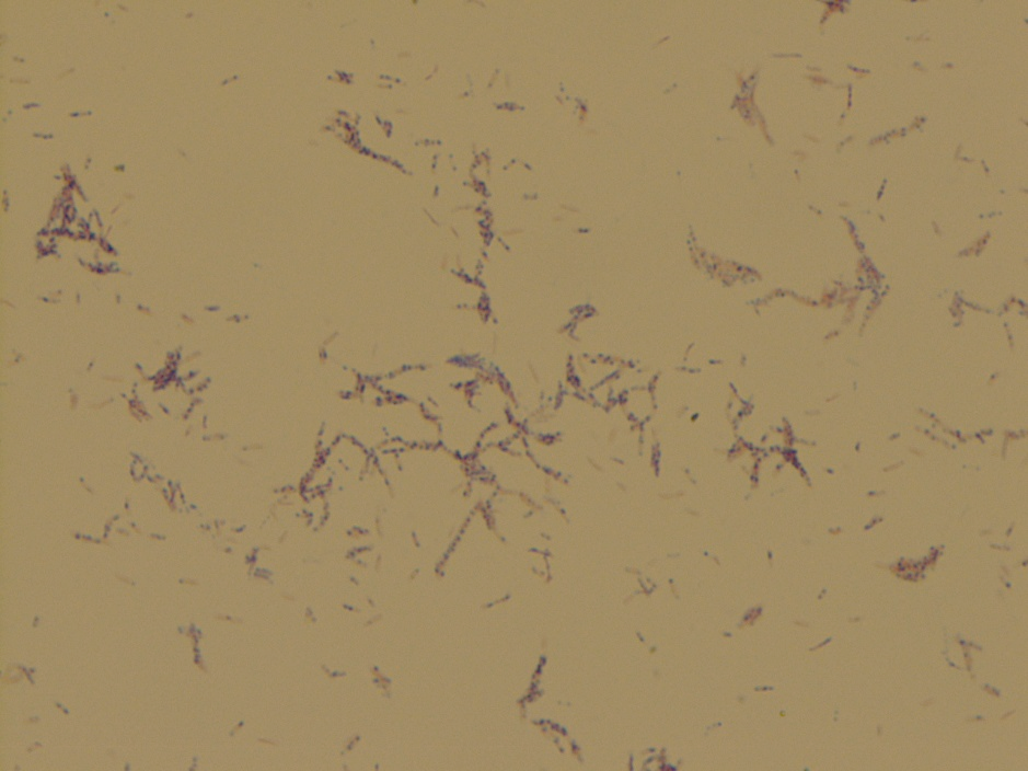
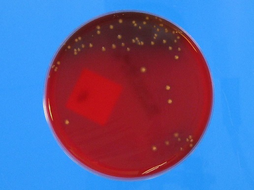
The stat gram stain showed many polymononuclear cells with moderate gram positive bacilli in a background of dark inorganic material (Image 1). Following 48 hours of incubation, there was anaerobic growth on the kanamycin and vancomycin (KV) and schaedler agar plates. A Gram stain of the broth showed gram positive bacilli arranged singly and in chains with some decolorization (Image 2). The KV and schaedler plates showed moderate growth of a single organism consisting of small glossy tan colored colonies (Images 3-4). No aerobic growth was observed on the blood, MacConkey, Columbia Naladixic Acid (CNA), or chocolate agar plates. Mass spectrometry (MALDI-TOF) identified the pathogenic organism as Clostridium innocuum.

Image 1. Synovial fluid Gram stain of the left hip showed moderate gram positive bacilli and many polymononuclear cells in a background dark inorganic debris (100x oil immersion).
Image 2. Gram stain from a positive broth culture showed gram positive bacilli arranged singly and in chains with some decolorization (100x oil immersion).
Image 3. Anaerobic growth on the schaedler agar showed growth of a single organism consisting of small round glossy tan colored colonies.
Image 4. Anaerobic growth on the kanamycin and vancomycin (KV) agar showed growth of a single organism consisting of small glossy tan colored colonies.

Discussion

Bacterial joint infections are more common in prosthetic joints as compared to native joints with a prevalence of 1-2% following hip arthroplasty (1). Most cases of bacterial septic arthritis are due to staphylococci (40 percent), streptococci (28 percent) or gram negative bacilli (19 percent) organisms (2). Joint infections secondary to anaerobes are less likely and account for 2-3% of all cases (3). A review of the literature shows less than 50 documented cases of septic arthritis due to Clostridium species. Amongst these cases Clostridium perfringens is the most commonly isolated pathogen (4). To date there are no documented cases of joint infections secondary to Clostridium innocuum species.

Clostridium innocuum is a non-motile, anaerobic, gram positive organism that reproduces by sporulation. These organisms are normally found as a part of the usual human gut flora and are rarely human pathogens. The name “innocuum” is derived from the term “innocuous” to convey the innocence of these organisms as they do not produce clostridial exotoxins. A review of the literature shows fewer than 20 reported cases of Clostridium innocuum infections with most reported cases being described in immunocompromised patients such as those with diabetes mellitus, chronic hepatitis, acquired immune deficiency syndrome (AIDS), leukemia, and organ transplantation (5-6). Clinically patients can present with a spectrum of symptoms which include fever of unknown origin, diarrhea/constipation, and non-specific respiratory symptoms. In almost all cases bacteremia ensued. Most cases were associated with a traumatic penetrating injury with few reported cases due to hematogenous spread (5-6).

Laboratory identification of Clostridium innocuum can be challenging due to its variable gram staining morphology and atypical colony morphology on differing culture media. Most traditional phenotypic methods can only reliably identify these organisms to the genus level as a Clostridium species. However, using mass spectrometry (MALDI-TOF) these organisms can be identified to the species level. Rapid identification of Clostridium innocuum from the subset of Clostridium species is clinically important as these organisms are the only known Clostridium species with intrinsic resistance to vancomycin (7). Although they do not possess clostridial exotoxins, these organisms are thought to have a lipopolysaccharide-like virulence factor and have a mortality rate comparable to toxigenic Clostridium species (7). Due to resistance to vancomycin, metronidazole, piperacillin and ampicillin-sulbactam are the alternative recommended first-line treatment options.

For this patient, following the results of the gram smear the patient was started on IV vancomycin but due to an adverse allergic reaction was switched to intravenous pencillin G and oral ciprofloxacin. He was subsequently taken to the operating room for incision and drainage and left hip revision arthroplasty with cup exchange. Blood cultures were collected post-operatively and showed no growth, possibly due earlier antibiotic administration. Susceptibility studies from Mayo Laboratories showed pan susceptibility to penicillin, piperacillin-tazobactam, ertapenem, clindamycin, and metronidazole. The patient was subsequently switched to intravenous penicillin and continued to show clinical improvement during his remaining hospital course.

References

  1. Horowitz DL, Katzap E, Horowitz S, Barilla-labarca ML. Approach to septic arthritis. Am Fam Physician. 2011;84(6):653-60.
  2. Ryan MJ, Kavanagh R, Wall PG, Hazleman BL. Bacterial joint infections in England and Wales: analysis of bacterial isolates over a four year period. Br J Rheumatol. 1997;36(3):370-3.
  3. Shah NB, Tande AJ, Patel R, Berbari EF. Anaerobic prosthetic joint infection. Anaerobe. 2015;36:1-8.
  4. Gredlein CM, Silverman ML, Downey MS. Polymicrobial septic arthritis due to Clostridium species: case report and review. Clin Infect Dis. 2000;30(3):590-4.
  5. Leal J, Gregson DB, Ross T, Church DL, Laupland KB. Epidemiology of Clostridium species bacteremia in Calgary, Canada, 2000-2006. J Infect. 2008;57(3):198-203.
  6. Lee NY, Huang YT, Hsueh PR, Ko WC. Clostridium difficile bacteremia, Taiwan. Emerging Infect Dis. 2010;16(8):1204-10.
  7. Chia JH, Feng Y, Su LH, et al. Clostridium innocuum is a significant vancomycin-resistant pathogen for extraintestinal clostridial infection. Clin Microbiol Infect. 2017;23(8):560-566.

-Noman Javed, MD is a 3rd year anatomic and clinical pathology resident at the University of Vermont Medical Center.

-Christi Wojewoda, MD, is the Director of Clinical Microbiology at the University of Vermont Medical Center and an Associate Professor at the University of Vermont.

Microbiology Case Study: A Man with History of ALL Presents with Fever and Diarrhea

Clinical History

A man in his 40’s with a past medical history of acute lymphoblastic leukemia/lymphoma (in remission), multiple infections including bacteremia and pulmonary aspergillosis, presented to the hospital with fever and diarrhea. Over the course of his stay, he had worsening renal function and developed profound hypotension and shock, which prompted initiation of two vasopressors and high-dose steroids. Eventually he developed acute hypoxic respiratory failure, requiring intubation. Complete blood count demonstrated an absolute eosinophilia of 8.58 x10⁹/L (reference range 0.04-0.62 x10⁹/L). Imaging revealed bilateral pulmonary infiltrates and a pleural effusion. Respiratory culture with gram stain was ordered for his tracheal aspirate, which revealed few polymorphonuclear cells, many gram-negative rods, yeast, and larvae of Strongyloides stercoralis (Image 1A). Wet mounts of the tracheal aspirate revealed numerous larvae and a few eggs of S. stercoralis (Image 1B-C); many of the larvae were motile (Movie 1). Stool examination of ova and parasites (O & P) were positive for larvae. Given the burden of organisms and prior administration of steroids, he was diagnosed with severe strongyloidiasis, consistent with hyperinfection. Concurrent blood cultures grew Enterococcus faecalis and Stenotrophomonas maltophilia; the respiratory culture also grew S. maltophilia, and tracks from the migrating larvae were observed on respiratory culture bacterial media (Image 1D).

Image 1. Tracheal aspirate Gram stain with S. stercoralis larvae, 100x objective magnification (A). Wet mount of tracheal aspirate revealing larvae (B) and eggs (C), 40x objective magnification. Blood agar plate growing S. maltophilia in an abnormal pattern, indicating motile larvae tracking through the agar (D).

Discussion

Strongyloidiasis is a spectrum of clinical disease caused by the nematode Strongyloides stercoralis.1,2 Descriptions of acute infection have been described in other Lablogatory entries here,3,4 and the full lifecycle is described in detail on the CDC DPDx website.5

Severe strongyloidiasis includes the syndromes of hyperinfection and disseminated disease. Hyperinfection is when there is an elevated burden of the typical autoinfection cycle involving the lungs and GI-tract. Usually there is an antecedent immunosuppressive event, such as administration of corticosteroids. Within the GI-tract lumen, increased numbers of rhabditiform larvae transform into the infective filariform larvae, which traverse the GI mucosa, migrate to the lungs via bloodstream/lymphatics where they enter alveolar air spaces, then ascend the respiratory tract, and are coughed up by the host and swallowed to re-enter the GI tract. In the GI tract adult females lay eggs through parthenogenesis, which give rise to further rhabditiform larvae. In extreme cases of hyperinfection, adults can be found in the lungs, where they may also lay eggs. Finding eggs in respiratory specimens is unusual, and may be related to the burden of disease.6

Disseminated disease is when larvae can be found in any additional organs/organ systems, such as the central nervous system, kidneys, liver, adrenals, etc. Invasive sampling is not typically performed, and larvae can be observed at autopsy.

Laboratory diagnosis of S. stercoralis involves identification of rhabditiform larvae in stool O &P exam; the presence of adults or eggs in stool is rare. Rhabditiform larvae have short buccal cavities and an ovoid genital primordium structure midway through the body (Movie 2). O&P exams can be performed on other body fluids, such as sputum and CSF. Serology can be useful to identify past exposure, especially prior to initiating immunosuppressive therapeutics such as corticosteroids. A nonspecific finding can be observed, as in this case, in the complete blood cell count and differential. Relative and absolute eosinophilia can be found in patients with parasitic infections; therefore, it is reasonable to rule out parasitic infection in this subset of patients. In the case presented here, the absolute eosinophilia was likely due to a persistent S. stercoralis infection, since these nematodes can live in the human host for decades.

The treatment of choice for severe strongyloidiasis is oral ivermectin, though albendazole is an alternative therapy. In some instances, subcutaneous ivermectin administration may be used.7

Follow-up

Oral ivermectin was administered to treat the strongyloidiasis and antibiotics were administered to treat the bacterial infections. Over the coming days, serial tracheal aspirates continued to reveal many larvae and eggs, so therapy was escalated to subcutaneous ivermectin. Over the course of therapy, the patient developed a fungemia with Candida guilliermondii. Despite aggressive antimicrobial therapy and intensive care, the patient remained hypoxemic and hypotensive. The family decided to transition to comfort measures and the patient passed away.

References

  1. Maguire JH. Intestinal Nematodes (Roundworms), in Mandell, Douglas, and Bennett’s Principles and Practice of Infectious Diseases, B. Mandell, Dolin, Editor. 2010, Elsevier: Philadelphia, PA. p. 3577-3586.
  2. Parasitology, in Koneman’s Color Atlas and Textbook of Diagnostic Microbiology, Procop et al., Editors. 2017, Lippincott Williams & Wilkins: China. p. 1452-1454.
  3. Kaur J, Stempak L. An 81 Year Old Female with Persistent Fevers. Lablogatory 2019 [cited 2019 11/5/2019]; Available from: https://labmedicineblog.com/2019/04/23/microbiology-case-study-an-81-year-old-female-with-persistent-fevers/.
  4. Mohammed M, Wojewoda C. A 47 Year Old Male with Abdominal Pain and Diarrhea. Lablogatory 2016 [cited 2019 11/5/2019]; Available from: https://labmedicineblog.com/2016/05/16/microbiology-case-study-a-47-year-old-male-with-abdominal-pain-and-diarrhea/.
  5. Centers for Disease Control. Strongyloidiasis. DPDx 2019 [cited 2019 11/5/2019]; Available from: https://www.cdc.gov/dpdx/strongyloidiasis/index.html.
  6. Keiser PB and Nutman TB. Strongyloides stercoralis in the Immunocompromised Population. Clin Microbiol Rev, 2004. 17(1): p. 208-17.
  7. Hurlimann E and Keiser J, A single dose of ivermectin is sufficient for strongyloidiasis. Lancet Infect Dis, 2019. 19(11): p. 1150-1151.

-IJ Frame, MD, PhD, Microbiology Fellow, University of Texas Southwestern Dallas, Texas

-Clare McCormick-Baw, MD, PhD is an Assistant Professor of Clinical Microbiology at UT Southwestern in Dallas, Texas. She has a passion for teaching about laboratory medicine in general and the best uses of the microbiology lab in particular.

Microbiology Case Study: 77 Year Old Man With History of Travel to India

Case Report

A 77 year old male presented to the hospital with chest pain, lightheadedness, burning urination for the past few weeks. He has blood in his urine due to a previously diagnosed neoplasm. The patient moved from India to the United States in February, with a diagnosis of bladder cancer and a history of hypertension, congestive heart failure, coronary artery disease, and atrial fibrillation. In the hospital, abscesses on both right and left kidneys were found, and patient had nephrostomy tubes placed. Purulent discharge confirmed he had a severe urinary tract infection.

Laboratory Identification

The patient’s urine culture grew >100,000 colony forming units/milliliter (CFU/ml) of an oxidase-positive, non-lactose fermenting Gram-negative rod. On the blood agar plate, large gray, smooth, flat, mucoid, β-hemolytic colonies were found. Although bacteria growing on solid media should not be actively smelled, the organism emitted a grape or tortilla smell from the plate. The organism was identified as Pseudomonas aeruginosa by MALDI-TOF mass spectrometry. The isolate was plated onto Mueller Hinton agar for Kirby-Bauer disc diffusion antibiotic susceptibility testing (Image 1). A fluorescent green lawn of bacteria grew up to the edge of all discs, indicating high-level resistance to all antibiotics tested (Table 1). Modified carbapenem inactivation method (mCIM) testing was positive and Cepheid GeneXpert CarbaR PCR testing revealed that this P. aeruginosa isolate carried the New Delhi metallo-β-lactamase-1 carbapenemase (NDM-1).

Image 1. Kirby-Bauer disc diffusion was used for antimicrobial susceptibility testing. Note no zones around any of the antibiotic discs, indicating resistance to all antimicrobials tested.
Table 1. Antimicrobial susceptibility testing interpretations. All drugs tested were resistant to this P. aeruginosa isolate.

Discussion

The issue of super bugs is on the rise, with the fear of antibiotic resistance disseminating through more bacterial populations and species. Carbapenems are drugs that are very powerful broad-spectrum antibiotics, usually reserved as a last resort treatment for serious and resistant infections.1 β-lactamases are divided into four Ambler classes: A, B, C, and D. Class B differs from the others because it utilizes zinc as a metal cofactor for its catalytic activity. The others use a serine residue for their catalytic activity.2

NDM-1 is a class B β-lactamase. It was named after New Delhi, India when a Swedish resident presented with an extremely resistant infection after a trip to India in 2008. NDM-1 bacteria can now be found with high prevalence in India and China, and increasingly in other countries such as the UK and US.3,4 While the origination of the gene may not have been India, many of these infections are from people who have traveled to India or other Asian continents.5 Concerns about overprescribing and misuse of antibiotics in India are rising, where India is one of the biggest consumers of antibiotics in the world. One study even found striking evidence of this misuse, demonstrating that 2 out of 3 adults under 20 presented antibiotic resistance isolates to fluoroquinolones and/or cephalosporins.6,7,8

Image depicting the NDM-1 protein anchored in the outer membrane of the bacterium. (Taken from Bahr, Guillermo, et al. “Clinical Evolution of New Delhi Metallo-β-Lactamase (NDM) Optimizes Resistance under Zn(II) Deprivation.” Antimicrobial Agents and Chemotherapy, vol. 62, no. 1, 2017, doi:10.1128/aac.01849-17.)

The gene for NDM-1 is blaNDM-1 and has been found on both plasmid and chromosomal components of different bacteria. Due to its presence on plasmids, the gene can easily spread through bacterial populations and other bacterial species – as has already been documented in Enterobacteriaceae and A. baumannii.3 The β-lactamase that it codes for is a lipoprotein that is anchored in the outer membrane of the gram negative bacteria. Other metalo-β-lactamases (MBLs) are periplasmic proteins, which are more affected by changes in essential metal cofactors in their enzymatic function. Thus far, it has been found that there are 16 discovered variants of NDM. Some variants being more fit than NDM-1. It is hypothesized that these variants are being selected for in the clinical setting, with the protein being more stable and demonstrating higher affinities for zinc during time of metal-chelating (a process the immune system adapts to combat infections).9 Unfortunately, NDM-1 and its variants are resistant to almost all antibiotics. Usually the only option is colistin and tigecycline.3

The disturbing issue, and the big picture, is the capability of MDR organisms and their genes of disseminating. As previously mentioned, NDM-1 is capable of spreading to other species and within its population. Yet, a terrifying report has demonstrated blaNDM-1 detection in artic soil samples from 2013, 4 years after the first detection of the gene.10 This demonstrates the ability for antibiotic resistance to spread on a global scale, and how serious this battle truly is.

References

  1. “Carbapenem-Resistant Enterobacteriaceae (CRE) Infection.” Centers for Disease Control and Prevention, Centers for Disease Control and Prevention, 23 Feb. 2015, http://www.cdc.gov/hai/organisms/cre/cre-patientfaq.html.
  2. Walther-Rasmussen, Jan, and Niels Høiby. “Class A Carbapenemases.” Journal of Antimicrobial Chemotherapy, vol. 60, no. 3, 2007, pp. 470–482., doi:10.1093/jac/dkm226.
  3. Khan, Asad U., et al. “Structure, Genetics and Worldwide Spread of New Delhi Metallo-β-Lactamase (NDM): a Threat to Public Health.” BMC Microbiology, vol. 17, no. 1, 2017, doi:10.1186/s12866-017-1012-8.
  4. Mohapatra P. R. (2013). Metallo-β-lactamase 1–why blame New Delhi & India?. The Indian journal of medical research137(1), 213–215.
  5. Roos, Robert. “Canada Finds More Infections with NDM-1 Resistance Factor.” University of Minnesota, Center for Infectious Disease Research and Policy, 11 Nov. 2010, http://www.cidrap.umn.edu/news-perspective/2010/11/canada-finds-more-infections-ndm-1-resistance-factor.
  6. Gupta, M., Didwal, G., Bansal, S., Kaushal, K., Batra, N., Gautam, V., & Ray, P. (2019). Antibiotic-resistant Enterobacteriaceae in healthy gut flora: A report from north Indian semiurban community. The Indian journal of medical research, 149(2), 276–280. doi:10.4103/ijmr.IJMR_207_18
  7. Kotwani, Anita, and Kathleen Holloway. “Access to Antibiotics in New Delhi, India: Implications for Antibiotic Policy.” Journal of Pharmaceutical Policy and Practice, vol. 6, no. 1, 2013, doi:10.1186/2052-3211-6-6.
  8. Kotwani, Anita, et al. “Antibiotic-Prescribing Practices of Primary Care Prescribers for Acute Diarrhea in New Delhi, India.” Value in Health, vol. 15, no. 1, 2012, doi:10.1016/j.jval.2011.11.008.
  9. Bahr, Guillermo, et al. “Clinical Evolution of New Delhi Metallo-β-Lactamase (NDM) Optimizes Resistance under Zn(II) Deprivation.” Antimicrobial Agents and Chemotherapy, vol. 62, no. 1, 2017, doi:10.1128/aac.01849-17.
  10. Mccann, Clare M., et al. “Understanding Drivers of Antibiotic Resistance Genes in High Arctic Soil Ecosystems.” Environment International, vol. 125, 2019, pp. 497–504., doi:10.1016/j.envint.2019.01.034.

-Ben Dahlstrom is a recent graduate of the NorthShore University HealthSystem MLS program. He currently works as a molecular technologist for Northwestern University in their transplant lab, performing HLA typing on bone marrow and solid organ transplants. He graduated with a bachelors in Biology at the University of Illinois at Chicago (UIC) and concurrently from the UIC Honors College. He discovered his passion for the lab through his experience in healthcare. His interests include microbiology, molecular, immunology, and blood bank.

-Erin McElvania, PhD, D(ABMM), is the Director of Clinical Microbiology NorthShore University Health System in Evanston, Illinois. Follow Dr. McElvania on twitter @E-McElvania. 

Microbiology Case Study: A 50 Year Old Male with Fevers

Case History

The patient is a 50 year old male who presented to urgent care with 5 days of fevers, chills, and myalgias. He reports no known tick bites, or prior treatment for tickborne illness, he travels frequently for work and has been in Pittsburgh, Omaha, Philadelphia, Charlotte, and Long Island over the past 3 months and is frequently outside golfing in Vermont and while traveling. At urgent care he had a CBC, CMP and urinalysis. His CBC was remarkable for leukopenia with absolute neutropenia (WBC count 2,790; Absolute neutrophil count 390) and thrombocytopenia (platelet count 26,000/cmm). His CMP was remarkable for a mildly elevated AST (66U/L) and total bilirubin (2.4mg/dL). A peripheral blood smear was made which revealed ring forms in his red blood cells. BINAX testing for malaria was negative. The next day he presented to the emergency department with left upper quadrant abdominal pain, night sweats, fatigue, fever, and blood in his urine where is was informed of his CBC results and was immediately started on azithromycin and atovaquone. Given the severity of his presentation a co-infection with Anaplasma phagocytophilum (Anaplasmosis) was suspected though the PCR testing was negative.

Laboratory Identification

Image 1. Giemsa stain of thin blood smears showing intracellular (left) and extracellular (right) ring-form organisms.

Thick and thin blood smears (recommended) were prepared which showed both intra and extracellular organisms with in normal sized red blood cells. BINAX testing for malaria was negative. Given the appearance and presence both within and outside the cell a diagnosis of Babesiosis was made.

Discussion

Babesiosis in the United States is caused by the microscopic parasite Babesia microti. Occasional cases caused by other species of Babesia have been detected (1). Babesia microti is spread by Ixodes scapularis ticks (also called blacklegged ticks or deer ticks) (1,2). Transmission mainly occurs in parts of the Northeast and upper Midwest; and it usually peaks during the warm months (1). Because Babesia shares a vector and geographic distribution with Borrelia burgdorferi (Lyme disease) coinfection of ticks with these two organisms can be seen in up to 40% of ticks and though the incidence of Babesiosis is much lower; up to 10% of Connecticut patients with seropositivity for Lyme disease are also seropositive for Babesia (2).

Most infections are probably asymptomatic. Manifestations of disease include fever, chills, sweating, myalgias, fatigue, hepatosplenomegaly, and hemolytic anemia. Symptoms typically occur after an incubation period of 1 to 4 weeks, and can last several weeks. The disease is more severe in patients who are immunosuppressed, splenectomized, and/or elderly (1,3).

During a blood meal, a Babesia-infected tick introduces sporozoites into the human host (1,3). Sporozoites enter erythrocytes and undergo asexual replication (budding). Multiplication of the blood stage parasites is responsible for the clinical manifestations of the disease (1). Humans are dead-end hosts and there is probably little, if any, subsequent transmission that occurs from ticks feeding on infected persons (1). However, human to human transmission is well recognized to occur through blood transfusions (1). Diagnosis is often rendered by direct visualization on thick and thin blood smears. Because the percent parasitemia is often low, the organisms can be easily missed, especially by automated hematology analyzers (3). Molecular methods such as PCR can be performed (3), but are not recommended as the first line test as the blood parasite exam with thick and thin blood smears are clinically sensitive in patients with symptomatic disease.

Most mild cases of Babesiosis will resolve spontaneously without treatment, especially in patient with a spleen (1,3). Treatment for more severe disease includes either azithromycin and atovaquone or clindamycin and quinine (1,3). If patients are severely immunocompromised and/or splenectomized can be treated with exchange transfusion in addition to antimicrobials (3).

References

  1. Centers of Disease Control and Prevention: Babesiosis. https://www.cdc.gov/parasites/babesiosis/
  2. Procop, Gary W., et al. Konemans Color Atlas and Textbook of Diagnostic Microbiology. 7th ed., Wolters Kluwer Health, 2017.
  3. Tille, Patricia M. Bailey & Scotts Diagnostic Microbiology. 13th ed., Elsevier, 2014.

-Casey Rankins, DO, is a 3rd year Anatomic and Clinical Pathology resident at the University of Vermont Medical Center.

-Christi Wojewoda, MD, is the Director of Clinical Microbiology at the University of Vermont Medical Center and an Associate Professor at the University of Vermont.

Microbiology Case Study: A 61 Year Old Male with Fevers and Weight Loss

Case History

The patient is a 61 year old male in good health until about 4 weeks prior to presentation when he sustained a tick bite on his left arm. He subsequently developed chills, fatigue, loss of appetite, and weight loss. Concerned that his symptoms were not improving, the patient presented to urgent care and a CBC was ordered. His CBC was remarkable for mild anemia (RBC count 3.96, HB 12.9) and thrombocytopenia (platelet count 78,000/cmm). Review of the peripheral blood smear revealed organisms present within his neutrophils. Given his history of a tick bite, Doxycycline was initiated for 14 days with immediate improvement of his symptoms, including a notable increase in appetite over the next few days.

Laboratory Identification

Image 1. Giemsa Stain showing a morulae within a neutrophil.

Within the neutrophils are purple organisms distinct from the nuclei identified as morulae. PCR testing for Anaplasma confirmed the result.

Discussion

 Anaplasmosis is a disease caused by the bacterium Anaplasma phagocytophilum, previously known as Ehrlichia phagocytophilum causing human granulocytic ehrlichiosis (HGE). A taxonomic change in 2001 identified that this organism belonged to the genus Anaplasma, and resulted in a change in the name of the disease to Anaplasmosis (1). These bacteria are obligate intracellular organisms in the Rickettsia family (1,2). Anaplasma cannot survive outside the cell and once it has been released, it rapidly induces uptake signals in other host cells (3). The number of Anaplasmosis cases reported to CDC has increased steadily since the disease became reportable, from 348 cases in 2000, to 5,762 in 2017(1).

Anaplasmosis is spread to people by tick bites primarily from the blacklegged tick (Ixodes scapularis) and the western black legged tick (Ixodes pacificus)(1,2). Anaplasmosis can be transmitted through blood transfusion and has been found in refrigerated blood more than a week after collection. Transfusion related infections have occurred from asymptomatic donors (1).

Signs and symptoms of Anaplasmosis typically begin within 1–2 weeks after the bite of an infected tick, which can be painless and often goes unnoticed. Early signs and symptoms (days 1-5) are usually mild or moderate and may include fever, chills, headache, muscle ache, nausea, vomiting, and lack of appetite (1,3). Rarely, if treatment is delayed or if there are other medical conditions present, Anaplasmosis can cause severe illness. Signs and symptoms of severe (late stage) illness can include respiratory failure, bleeding problems, organ failure, and death. Laboratory findings can include mild anemia, thrombocytopenia, leukopenia (characterized by relative and absolute lymphopenia and a left shift) and mild to moderate elevations in hepatic transaminases (1). Abnormal laboratory findings can appear in the first week of illness; however, normal laboratory findings do not rule out possible infection.

Co-infection with other tick borne illnesses such as Borrelia burgdorferi (Lyme disease), Babesia microti (Babesiosis), Ehrlichia muris eauclairensis (Erlichiosis), and Powassan virus can be seen so additional testing may be necessary in some patients. Methods for diagnosing Anaplasmosis include serology, molecular methods, and morphological identification. Though morphologic identification is extremely specific is lacks sensitivity making molecular methods such as PCR the diagnostic methods of choice (2). Treatment for most Rickettsial illnesses including Anaplasmosis are tetracyclines, especially doxycycline which is the drug of choice (1,3).

References

  1. Centers for Disease Control and Prevention: Anaplasmosis. https://www.cdc.gov/anaplasmosis/index.html
  2. Procop, Gary W., et al. Konemans Color Atlas and Textbook of Diagnostic Microbiology. 7th ed., Wolters Kluwer Health, 2017.
  3. Tille, Patricia M. Bailey & Scotts Diagnostic Microbiology. 13th ed., Elsevier, 2014.

-Casey Rankins, DO, is a 3rd year Anatomic and Clinical Pathology resident at the University of Vermont Medical Center.

-Christi Wojewoda, MD, is the Director of Clinical Microbiology at the University of Vermont Medical Center and an Associate Professor at the University of Vermont.

Microbiology Case Study: An 18 Year Old with Gastrointestinal Bleeding

An 18 year old female with no significant past medical history experienced multiple episodes of gastrointestinal bleeding over the course of a few weeks. The most recent bout included a bloody episode that filled the toilet, for which she provided a picture for the clinician. She denies any other associated symptoms including epigastric pain, nausea, vomiting, fever, or chills. Her travel history is unknown.

Review of her history reveals an unremarkable family and social history. She has never had an incident similar to this in the past and no other family members have ever complained of similar symptoms. Review of systems was unremarkable and within normal limits. Physical exam was unremarkable. A rectal exam was performed and was noted to have brown stool that was guaiac (occult blood) positive. Non bleeding internal hemorrhoids were noted. There were no external hemorrhoids present.

Labs drawn including CBC were within normal ranges with the exception of absolute eosinophils which were at the upper limit of normal range at 0.6 x 10³/µL [normal range= 0.0 – 0.6 103/µL].

The patient had an esophagogastroduodenoscopy (EGD) to further investigate the gastrointestinal bleed. The exam was otherwise normal with exception of the ascending colon where they noted a worm on the surface of the mucosa (Image 1-2). The worm was collected and transported to microbiology for examination (Image 3-4).

Image 1. View of a worm seen on the mucosal surface of the ascending colon.
Image 2. Another view of a worm seen on the mucosal surface of the ascending colon.
Image 3. Adult worm viewed under the dissecting microscope.
Image 4. Eggs viewed under the dissecting microscope.

Discussion

Examination of the worm and eggs revealed morphology consistent with Trichuris trichiura, or whipworm.

T. trichiura is most prevalent in warm, moist regions. The worldwide prevalence of infection is estimated to be roughly 800 million, mostly among poorer populations. Infection from T. trichiura is spread via fecal-oral route and caused by ingesting embryonated eggs. This occurs when contaminated dirt is ingested or by consumption of vegetables or fruits that have not been carefully cooked, washed or peeled.

The male and female worms both have the long whip-like structures at the anterior end. T. trichiura worms are 30-50 mm in length and the average life span is 1 year but they can live up to 10 years. The females have a straight and thick head while the males have a curly ended head. The males are typically longer the females. The eggs classically have barreled shaped, brown eggs with thick shells that measure 50-55 µm long by 22-24 µm wide. At each pole is lucent mucoid plug. The can also vary in size as noted in Image 5.

The adult female T. trichiura produces 1,000-7,000 eggs per day. The life cycle begins as unembryonated eggs passed in feces into soil (Figure 1). It takes approximately 21 days in the soil for an unembryonated egg to go through the process of embryonation to become the infective form of the parasite. Once ingested, the embryonated eggs hatch in the human intestine.

Image 5. T. trichiura eggs (CDC DPDx website)
Figure 1. Lifecycle of T. trichiura (CDC, DPDx)

Clinically, symptoms vary depending on the worm biomass present with most infections being asymptomatic. Symptoms include cramping, weight loss, growth restriction in children, bloody stool, and anemia. It can also result in Trichuris dysentery syndrome, which is more common in children. Recurrent rectal prolapse has also been reported. Lab findings include peripheral eosinophilia. T. trichiura is treated with Albendazole for 5-7 days +/- Ivermectin. Our patient was then prescribed albendazole and is being followed in GI clinic.

References

  1. Centers for Disease Control and Prevention. “Laboratory Identification of Parasites of Public Health Concern: Trichuriasis”. https://www.cdc.gov/dpdx/trichuriasis/index.html
  2. Procop, G. W., Church, D. L., Hall, G. S., Janda, W. M., Koneman, E. W., Schreckenberger, P. C., & Woods, G. L. (2017). Koneman’s color atlas and textbook of diagnostic microbiology (Seventh edition.). Philadelphia: Wolters Kluwer Health.

-Sharif Nasr, MD, 4th year anatomic and clinical pathology resident at University of Chicago (NorthShore). Dr. Nasr has an interest in GI pathology.

-Erin McElvania, PhD, D(ABMM), is the Director of Clinical Microbiology NorthShore University Health System in Evanston, Illinois. Follow Dr. McElvania on twitter @E-McElvania. 

Microbiology Case Study: A 70 Year Old Male with Multiple Myeloma

Case History

The patient is a 70 year old male who was diagnosed with Kappa free light chain multiple myeloma. He was initially seen after he had a fall in the woods and underwent imaging which showed multiple lytic lesions and blood work showing monoclonal proteins and thrombocytopenia. He was found to have a lesion on his right scapula for which he received radiation. Bone marrow biopsy was performed which showed 60% plasma cells. To date he has completed radiation therapy, 5 cycles of chemotherapy, and is in the process of collecting stem cells for autologous stem cell transplant. Routine fungal culture of the stem cell collection grew a single tan white dry appearing colony on potato flake agar. A Gram stain of the organism revealed gram positive cocci mixed with filamentous structures.

Laboratory Identification

Image 1. Single tan white dry colony on potato flake agar.
Image 2. Modified acid fast stain (left) and Gram stain (right).
Image 3. Filamentous branching on Gram stain.

Based on the colony morphology and Gram stain results the organism was suspected to be in the Streptomyces genus. Identification with MALDI-TOF was attempted and did not yield a result as this bacteria is not in the data base.

Discussion

Streptomyces is a genus of gram positive aerobic saprophytic bacteria that grows in various environments, and has a filamentous form similar to fungi (1). The morphologic differentiation of Streptomyces involves identification of complex multicellular architecture with germinating spores that form hyphae, and multinuclear aerial mycelium, which forms septa at regular intervals, creating a chain of uninucleated spores (2,3). They are able to metabolize many different compounds including sugars, alcohols, amino acids, and aromatic compounds by producing extracellular hydrolytic enzymes (helping with degradation of organic matter). Their metabolic diversity is due to their extremely large genome which has hundreds of transcription factors that control gene expression, allowing them to respond to specific needs (3).

Streptomyces is also considered to be one of the most medically important bacteria because of its ability to produce bioactive secondary metabolites. These metabolites are used in the creation of antifungals, antivirals, antitumoral, anti-hypertensives, and many antibiotics and immunosuppressives. They are responsible for 2/3 of all the worlds naturally occurring antibiotics (1).

Streptomyces is usually considered a laboratory contaminant though they can cause infections in immunocompromised patients and are chiefly responsible for granulomatous lesions in skin also known as actinomycotic mycetomas (1,2). Invasive pulmonary disease has been seen in HIV patients, splenectomized patients with sarcoid, and rarely in immunocompetent hosts (1). More rare presentations include brain abscesses can be seen in patients with cerebral trauma, peritoneal infections have been shown to occur in patients undergoing multiple pericenteses, and bacteremia in patients with indwelling catheters (1). Infection with Streptomyces is not common so susceptibility data is limited. Available data shows that organisms were consistently susceptible to amikacin; frequently susceptible to imipenem, clarithromycin or erythromycin, minocycline, and trimethoprim-sulfamethoxazole; and infrequently susceptible to ciprofloxacin and ampicillin (4).

Our patient had not received the stem cell unit that this grew from, so another aliquot was requested. The second aliquot did not grow any organisms, so the Streptomyces was considered a contaminant.

References

  1. Procop, Gary W., et al. Konemans Color Atlas and Textbook of Diagnostic Microbiology. 7th ed., Wolters Kluwer Health, 2017.
  2. Tille, Patricia M. Bailey & Scotts Diagnostic Microbiology. 13th ed., Elsevier, 2014.
  3. Chater KF. Recent advances in understanding Streptomyces. F1000Res. 2016;5:2795. Published 2016 Nov 30. doi:10.12688/f1000research.9534.1
  4. Mona Kapadia, Kenneth V.I. Rolston, Xiang Y. Han, Invasive Streptomyces Infections: Six Cases and Literature Review, American Journal of Clinical Pathology, Volume 127, Issue 4, April 2007, Pages 619–624, https://doi.org/10.1309/QJEBXP0BCGR54L15

-Casey Rankins, DO, is a 3rd year Anatomic and Clinical Pathology resident at the University of Vermont Medical Center.

-Christi Wojewoda, MD, is the Director of Clinical Microbiology at the University of Vermont Medical Center and an Associate Professor at the University of Vermont.

Microbiology Case Study: A 40 Year Old Woman with Fever, Chills, and Leg Pain

Clinical History

A 40 year old African American female with a history of sickle cell disease presented to an outpatient clinic with fever, chills, and leg and back pain consistent with a sickle cell crisis. Her past medical history was also significant for asthma and seizures. She rated her pain as 10 out of 10, her vitals showed a temperature of 101.0°F, and she was also tachycardic and hypotensive. Her white blood cell count was 23.0 TH/cm2, hemoglobin 8.4 g/dL, hematocrit 26.0%, and platelets 619,000 TH/cm2. In clinic, she received pain medications and a fluid bolus, two sets of blood cultures were collected, and she was transferred to the emergency department for further work up.

Laboratory Identification

Image 1. Gram stain from a positive blood culture bottle showing small, gram positive budding yeast (1000x oil immersion).
Image 2. A mucoid, salmon-colored yeast grew on Sabouraud dextrose and chocolate agars.

Blood culture bottles were positive after approximately two days on the automated instrument. The Gram stain showed small, gram positive budding yeast (Image 1). The BioFire FilmArray for blood culture identification was negative for Candida albicans, C. glabrata, C. krusei, C. parapsilosis, and C. tropicalis. At this time, she was started on micafungin for antifungal therapy. A mucoid, salmon colored yeast grew on both Sabouraud dextrose and chocolate agars (Image 2) and was identified by Vitek 2 as Rhodotorula spp.

Discussion

Rhodotorula spp. are basidiomycetous yeasts that make up the normal microbiota on moist skin and can be found in bathtubs and on shower curtains. Rhodotorula spp. are usually considered contaminants, but can rarely cause fungemia in patients with central lines, endocarditis, peritonitis, and meningitis, especially in those that are immunocompromised. R. mucilaginosa, R. glutinis, and R. minuta are the species commonly associated with human disease. 

In the laboratory, Rhodotorula spp. grow as a mucoid, salmon colored yeast within 1-3 days of incubation. On Gram stain or lactophenol cotton blue prep, the yeast is small and round to oval with multilateral budding. Pseudohyphae are not usually present. Rhodotorula spp. produce urease and fail to ferment carbohydrates. R. mucilaginosa is negative for nitrate assimilation. Identification can also be confirmed by commercial kits, automated systems, and MALDI-TOF mass spectrometry. Rhodotorula spp. are intrinsically resistant to echinocandins and fluconazole.

In the case of our patient, she was switched to intravenous amphotericin B after the identification of Rhodotorula spp. was made. Reference laboratory testing identified the isolate as R. mucilaginosa with high minimum inhibitory concentrations (MIC) to fluconazole and echinocandins. Amphotericin had an MIC of 0.5 µg/ml. She successfully completed a 14 day course with close monitoring of creatinine, electrolytes, and platelet count. Repeat blood cultures were negative and no other focuses of infection were found on CT scans, transthoracic echocardiogram, and ophthalmology exam.

-Lisa Stempak, MD, is an Assistant Professor of Pathology at the University of Mississippi Medical Center in Jackson, MS. She is certified by the American Board of Pathology in Anatomic and Clinical Pathology as well as Medical Microbiology. She is the Director of Clinical Pathology as well as the Microbiology and Serology Laboratories. Her interests include infectious disease histology, process and quality improvement, and resident education.

Microbiology Case Study: A 35 Year Old Female with Post-Op Drainage

Case History

A 35 year old female with a history of BRCA-positive breast cancer (status-post right radical mastectomy February 2018) underwent prophylactic left mastectomy and revision of right mastectomy. She received prophylactic clindamycin and cefazolin. She was discharged on post-op day 5 with bacitracin and 3 weeks of cefadroxil. She initially healed well. On post-op day 43 she noted drainage from the left incision site. At presentation she was afebrile.  There was a 3.0 x 2.0 cm area of induration and erythema on the right lateral aspect of her abdominal incision with seropurulent drainage. Incision and drainage was performed in office and a swab of the fluid was sent to microbiology. The initial gram stain and cultures were negative for bacteria. The patient was placed on sulfamethoxazole-trimethoprim and levofloxacin. At follow up 7 days later, another abscess was medial to the prior site was incised and drained. A swab of the fluid was sent to microbiology for bacterial and fungal cultures.

Laboratory Identification

Fungal cultures grew at 36 hours on potato-flake agar. Gram stain revealed gram-variable bacilli. Growth on 7H10 agar produced colonies at 72 hours, and Kinyoun staining was positive for acid-fast bacilli. Matrix assisted laser desorption ionization-time of flight mass spectrometry (MALDI-TOF) at 72 hours identified Mycobacterium abscessus complex.

Image 1. Growth on 7H10 agar.
Image 2. Gram stain from 7H10 agar showing gram variable bacilli.
Image 3. Kinyoun stain showing acid-fast bacilli.

Discussion

M. abscessus complex is a group of rapidly-growing, nontuberculous mycobacteria. As such they are acid-fast bacilli that grow within 7 days when transferred from solid media to solid media. The subspecies are M. abscessus abscessus, M. abscessus massiliense, and M. abscessus bolletii. The complex is known to cause progressive pulmonary disease in patients with underlying structural lung diseases. It has been estimated to comprise up to 13% of all mycobacterial pulmonary infections. It has also been implicated in skin and soft tissue infections (SSTIs) following surgical procedures or environmental exposure (i.e. spas). SSTIs can also occur by seeding from disseminated disease. Rarer manifestations include central nervous system (CNS) and ocular involvement. Identification is by culture and molecular techniques. It is classically resistant to many drug classes with limited consensus on appropriate therapy. It can harbor the erm gene, which confers inducible erythromycin resistance. Clarithromycin, amikacin, and cefoxitin tend to have the lowest rates of resistance. Long-term multidrug regimens are recommended, based on susceptibility testing. Changes to initial therapy are usually required due to side effects or lack of efficacy. Surgical therapy is often required, when possible. Mortality post therapy is approximately 15%.

At two-week follow up, the wound had no purulent drainage or erythema. The plan was for prolonged three-drug therapy tailored to susceptibility data.

  1. Griffith DE. Rapidly growing mycobacterial infections: Mycobacteria abscessus, chelonae, and fortuitum. Von Reyn CF and B A, eds. UpToDate. Waltham, MA: UpToDate Inc. https://www.uptodate.com (Accessed on May 21, 2019.)
  2. Lee MR, Sheng WH, Hung CC, Yu CJ, Lee LN, Hsueh PR. Mycobacterium abscessus Complex Infections in Humans. Emerg Infect Dis. 2015;21(9):1638–1646.
  3. Novosad SA, Beekmann SE, Polgreen PM, et al. Treatment of Mycobacterium abscessus Infection. Emerging Infectious Diseases. 2016;22(3):511-514.

-Jonathan Wilcock, MD is a 1st year anatomic and clinical pathology resident at the University of Vermont Medical Center.

-Christi Wojewoda, MD, is the Director of Clinical Microbiology at the University of Vermont Medical Center and an Associate Professor at the University of Vermont.

Microbiology Case Study: A 55 Year Old Male with Altered Mental Status

Case History

A 55 year old male presented to the emergency department (ED) with altered mental status (AMS). His past medical history includes stage 4 pancreatic cancer with known invasion into the distal splenic vein. Currently undergoing chemotherapy, his last infusion was one week prior to presentation. On physical exam, the patient is a cachectic male with dry mucous membranes, scleral icterus, hypotension (79/37), hypothermia (35o C), tachypnea (Respiratory Rate of 20/min) and tachycardia (pulse up to 130s). Initial labs were ordered including blood cultures and were significant for hypoglycemia (40mg/dL), pancytopenia, mild liver function test abnormalities, an ammonia level of 80 µmol/L and lactate of 11.2 mmol/L. It was concluded that the AMS resulted as a consequence of hypoglycemia. However, there was also concern for intracranial pathology vs. stroke vs. metastatic disease. Brain imaging showed no obvious lesion or mass. The cause of the hypotension was uncertain but could be a result of volume depletion or sepsis. Empiric vancomycin and cefepime were initiated in the ED. However, the patient decompensated, developing mid-abdominal and periumbilical ecchymoses suspicious for a retroperitoneal hemorrhage. Despite aggressive therapy, he expired in the ED.

Laboratory Identification

An anaerobic blood culture bottle flagged positive at 5 hours incubation. A gram stain showed large, boxy, gram positive rods without spores (Image 1). Brucella blood agar was inoculated and incubated anaerobically.  Following overnight incubation, the surface of the plate showed a subtle film of growth covering the plate and detectable hemolysis (Image 2). No discreet colonies were identified. A catalase test was performed and was negative. Definitive identification of Clostridium septicum was obtained by MALDI-TOF.

Image 1. Gram stain from the anaerobic blood culture bottle that flagged as positive following 5 hours of incubation. Boxy, large gram positive rods without spores are observed. Oil immersion photomicrograph (x100 objective).
Image 2. Brucella agar plate following 24 hours of incubation under anaerobic conditions at 35oC. Significant bacterial growth in a haze and detectable Beta hemolysis can be observed. No discreet colonies can be identified. Identification by MALDI-TOF was Clostridium septicum.

Discussion

Clostridium septicum is an anaerobic gram positive bacillus that can produce spores; however, spores are not frequently seen, especially in nutrient-rich environments. Spores, when present, are typically oval and located subterminally. Infection by C. septicum was once thought to be extremely rare, but improvements in anaerobic laboratory techniques have allowed for the discovery of the true potential of this agent. C. septicum is one of several bacteria that can cause myonecrosis (i.e., gas gangrene). Infections are typically seen in settings of immunodeficiency, trauma, surgery, malignancy, skin infections/burns, and septic abortions. The colon may promote the growth of C. septicum better than other anatomic sites due to its anaerobic conditions. As one of the more aggressive etiologies of gas gangreneC. septicum infection progresses very rapidly, with a mortality rate of approximately 79% in adults, typically occurring within 48 hours of infection. Symptoms of infection include pain, described as a heaviness or pressure that is disproportionate to physical findings, tachycardia, and hypotension. Tissue necrosis then causes edema and ischemia resulting in metabolic acidosis, fever, and renal failure. The carbon dioxide and hydrogen produced during the growth of the organism move through tissue planes, causing their separation, producing features characteristic of palpable emphysema (i.e., crepitus). This also results in a magenta-bronze skin discoloration and bulla filled with a foul-smelling serosanguinous fluid.

Four toxins have been isolated from C. septicum: the lethal alpha toxin, DNase beta-toxin, hyaluronidase gamma toxin, and the thiol-activated/septicolysin delta toxin. Alpha toxin causes intravascular hemolysis and tissue necrosis and is well known as the primary virulence factor of C. septicum

C. septicum derived gas gangrene has shown strong correlations with increased levels of malignancy. Patients with C. septicum infections may have an occult colon cancer or a tumor that has metastasized to the colon. C. septicum bacteremia is also associated with typhlitis (defined as inflammation of the cecum that can extend proximally into the terminal ileum or distally into the ascending colon), which can develop in patients with hematologic malignancy receiving chemotherapy. Because the organism may be harbored in the gastrointestinal tract, the organism may gain access to the bloodstream through the ileocecal region.

Therapy includes antibiotics and surgical debridement (with occasional amputation). For antibiotic selection, typical anaerobic coverage includes piperacillin/tazobactam, ampicillin/sulbactam, metronidazole or meropenem. Vancomycin is also effective. Susceptibility testing is not typically performed; moreover, the CLSI makes an annual antibiogram which can be used as a guide. 

Key points

  • C. septicum often has swarming growth that covers the plate surface.
  • Spontaneous myonecrosis with C. septicum bacteremia can be an indicator of possible occult colonic malignancy.
  • C. septicum can be associated with typhlitis in neutropenic patients with hematologic malignancy undergoing chemotherapy.

 References

  1. Smith-Slatas CL, Bourque M, and Salazar JC (2006). Clostridium septicum infections in children: a case report and review of the literature. Pediatrics 117(4): e796-e805.
  2. Alpern, RJ and Dowell, VR (1969). “Clostridium septicum infections and malignancy”. JAMA. 209: 385–388.
  3. Ballard, J, Crabtree, J, Roe, BA, and Tweten, RK (1995). “The primary structure of Clostridium septicum alpha-toxin exhibits similarity with that of Aeromonas hydrophila aerolysin”. Infection and Immunity. 63 (1):340–344.
  4. Sidhu JS, Mandal A, Virk J, and Gayam V (2019). “Early detection of colon cancer following incidental finding of Clostridium septicum bacteremia”. J Investig Med High Impact Case Rep. Jan-Dec;7:2324709619832050.
  5. Srivastava I, Aldape MJ, Bryant AE, and Stevens DL. (2017). “Spontaneous C. septicum gas gangrene: A literature review” Anaerobe. Dec;48:165-171

Xiang Xu, MD, PhD and Dominick Cavuoti, DO contributed to this case.

-Clare McCormick-Baw, MD, PhD is an Assistant Professor of Clinical Microbiology at UT Southwestern in Dallas, Texas. he has a passion for teaching about laboratory medicine in general and the best uses of the microbiology lab in particular.