Deep Dive into the Laboratory

This post is going to travel slightly off the beaten path by presenting a comparative analysis of my two favorite things – scuba diving and laboratory medicine. What in the world, you say? How can you possibly liken a recreational activity to an entire field of study? Easy. You just have to have a passion for both! I’ve been swimming and snorkeling since I was a wee little kid, always fascinated with being able to free dive down to the bottom of a body of water in one breath (you can blame my mermaid encounter for that). Around that same time, my parents gave me first microscope. The obsession with water was real, always examining wet mounts of everything that could fit between the slide and the coverslip. All throughout my grade school and undergrad years, I flourished in any science course that featured a microscope, finally acknowledging the fact that I had a knack for cells and tissues under the light microscope. During each summer break, I’d travel down to Florida with my parents and embrace the hidden gems of freshwater springs. It wasn’t until my junior year of college where I took a histology course during my summer break that I realized I wanted to make a career of this. During that summer, I began to research scuba diving; however, it wasn’t until after grad school and establishing a career as a cytologist that I finally committed to the idea. 10 years after graduating from a cytology program, I became the cytology supervisor at a renowned cancer center, and six years after my initial scuba diving certification, I became a dive professional, earning the title of divemaster.

Now, you should know that I like to dive deep into things – be it 112 feet down into a dark cavern or fully immersing myself in my work. Here, we’re going to dive into both scuba and the laboratory.

First and foremost, in both fields – diving and laboratory medicine, safety is paramount. There are rules and mechanisms in place to ensure that no one gets hurt. Policies and procedures are designed to keep you, your peers, and your “customers” (patients and students/certified divers) safe. Failure to follow a procedure can jeopardize lives. I’m certain you understand where I’m going with this. There are inherent risks in both fields, and we try to error-proof as best as we can. In diving, safety checks include checking you and your dive buddy’s equipment and configuration to make sure that gas cylinders are turned on and filled to an appropriate pressure, all hoses are connected and work as designed, releases and weights are secured, and that the divers have everything they need to perform the dive. Fortunately, in the laboratory, automation and barcoding has relieved the human eye of some of the burden of solely checking every patient detail, every order, and every equipment’s function. However, laboratory professionals must do their due diligence to double check. No piece of equipment is perfect, but following a procedure designed to avoid errors is the first step in verifying that the correct test has been ordered and the correct results will be delivered to the correct patient.

If you thought that preventative maintenance was necessary for lab equipment, scuba gear is the same way. Unlike a laboratory inspection, diving, for the most part, does not require evidence of gear maintenance to be “compliant” and therefore fit for use. The exception is visual inspection and hydrostatic testing of gas cylinders, but this is more a Department of Transportation rule. With that said, one of the top three reasons for dive accidents is equipment failure due lack of routine maintenance. That’s a risk I’m not willing to take. As much as I wish I had gills, I’m limited to the human form and the environment we are designed to live in. So whether it’s your own life or a family member’s test result, make sure that the relevant equipment is maintained per the manufacturer’s instructions or a more stringent policy or regulation.  

One of the many things I practice (while still ensuring a lean mindset) is redundancy. In diving, redundancy comes in the form of equipment. At a minimum, you should have two computers, two flashlights, two air sources, two regulators, two masks, two cutting implements, two people (buddy team), etc. If something goes wrong during a dive, there is a backup that can get you out of jam so that you can safely continue, or if necessary (and more likely the case), end that dive and formulate a plan for future dives. Think of needing to end the dive as a downtime procedure. In the laboratory, redundancy is having two centrifuges, two staining machines, two accessioning workstations, two analyzers (if that’s even in the budget), and other alternative, yet necessary procedures in the event of an electronic or equipment failure. The concept of redundancy is to maintain operations AND safety if something goes wrong.

Lastly, and something that I end my staff huddles or one-on-ones with is personal well-being. In diving, we disclose that it is okay to call a dive at any point for any reason, and a reason does not have to be provided. If you deem yourself physically, emotionally, or mentally unfit for a dive on any given day, call it. Do not muster through and pretend everything will be okay. That’s when divers neglect equipment checks, or fail to watch their gas consumption, depth limits, or time spent underwater. That’s when accidents happen. When you cannot be present for a dive, you cannot be present for your buddy or your students or divers that you are leading. In laboratory medicine, if you are not well, take the day off. I know we are dealing with staffing shortages across the country, I know that it might shift additional work onto a colleague, but you cannot risk jeopardizing patient care because you neglected yourself. Putting on a brave face for others in a world where it’s encouraged to be vulnerable is detrimental to more than just yourself, it’s detrimental to your passions. So take care of yourself and your (buddy) teams so that you can keep your passions safe and productive.

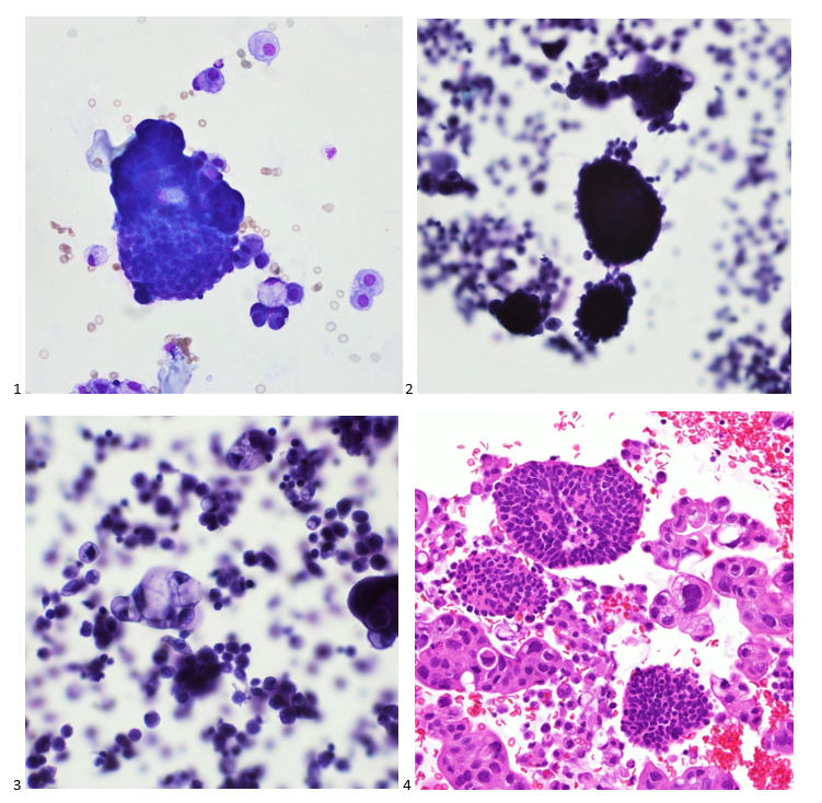
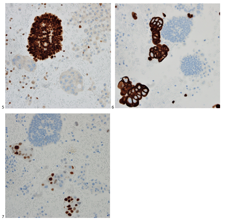
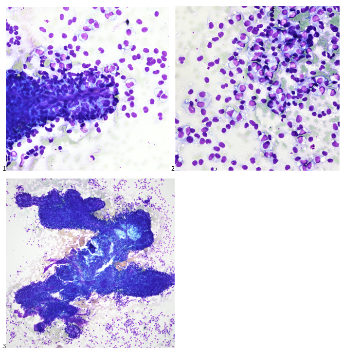
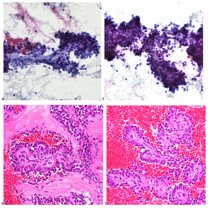
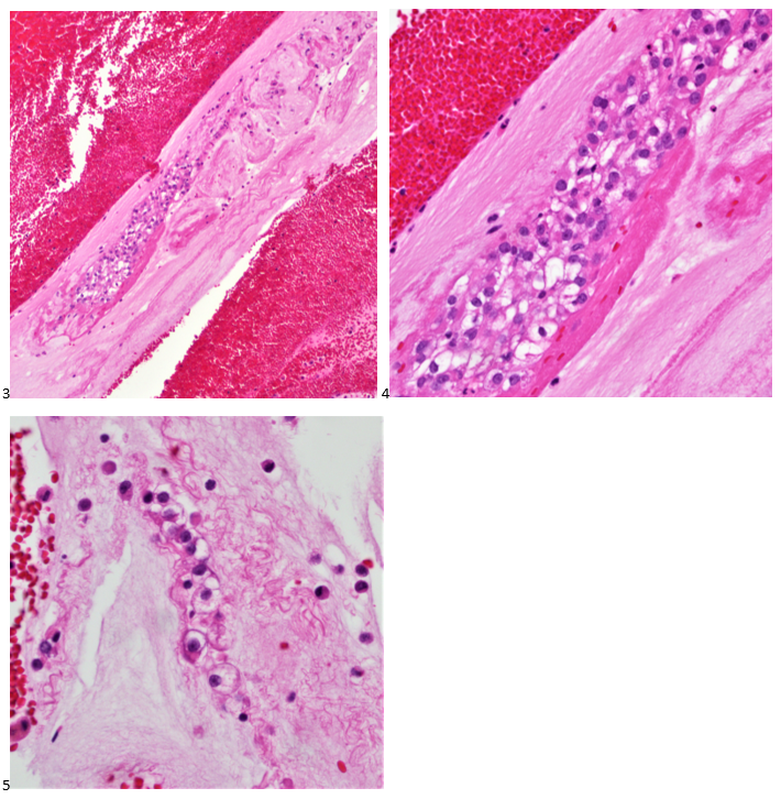
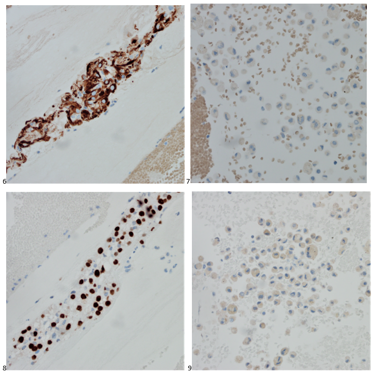
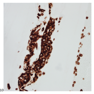
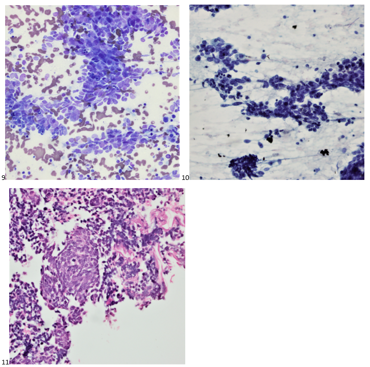
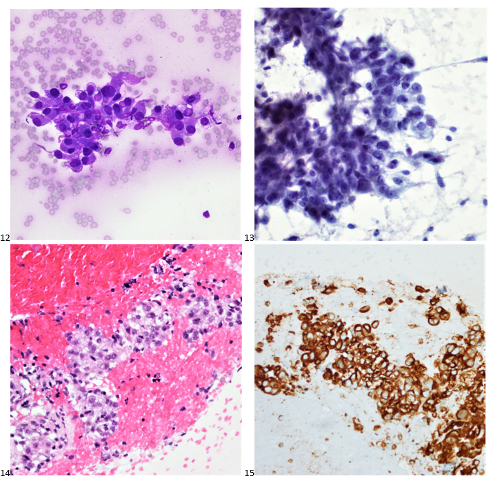
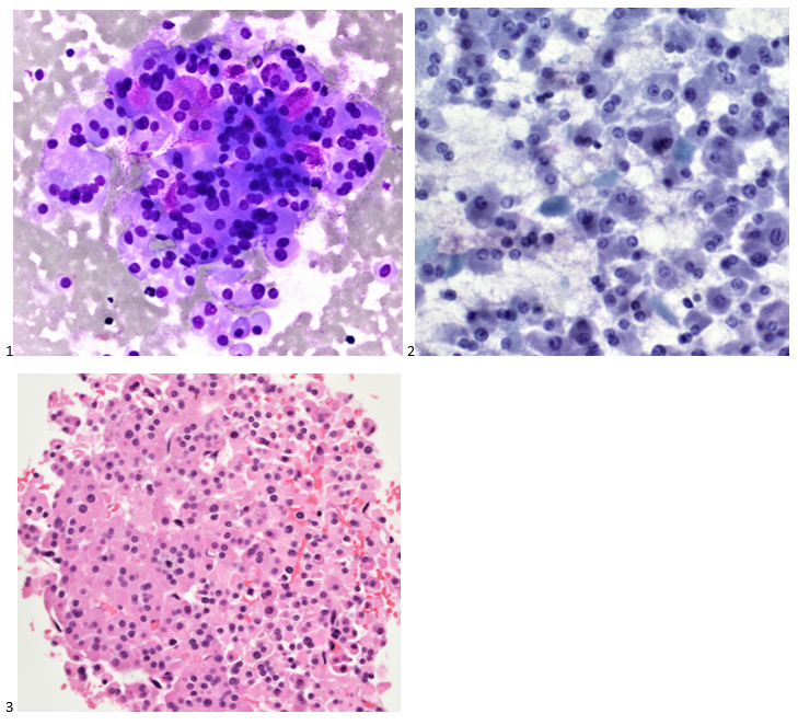
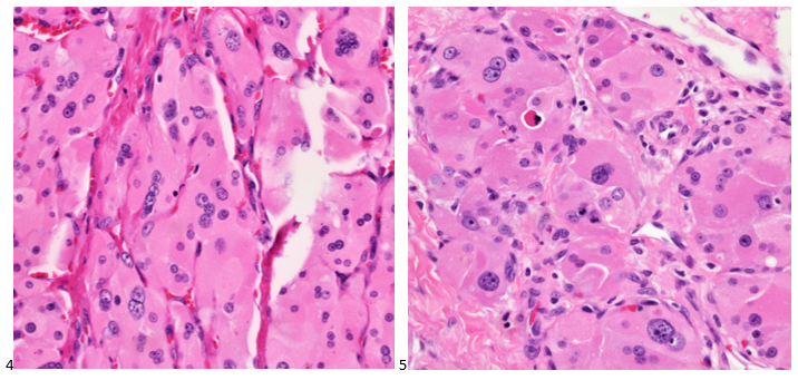
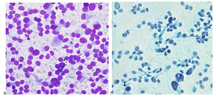
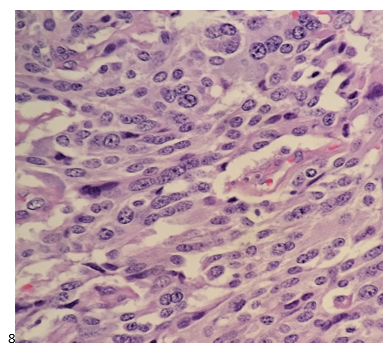
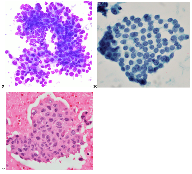
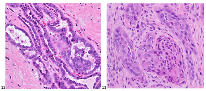
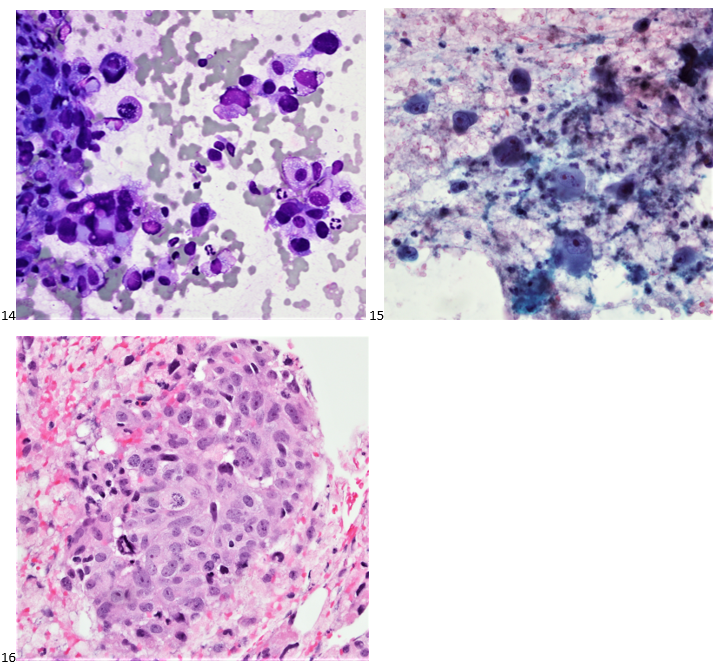
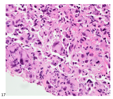
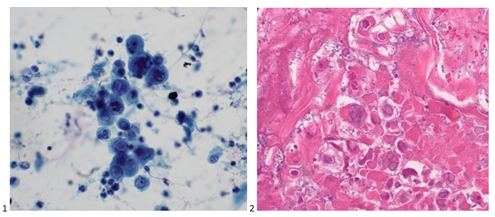
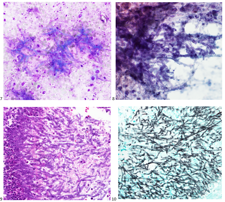
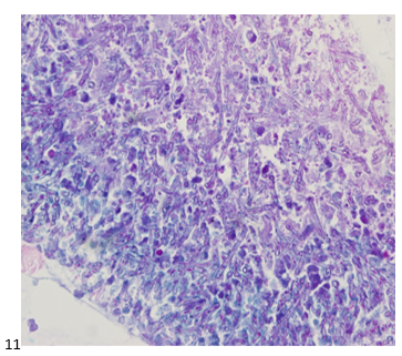
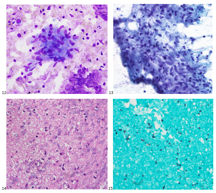
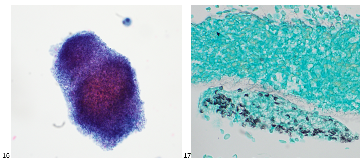
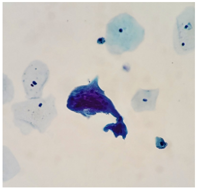
P.S. Sometimes diving and cytology overlap under the microscope, too!

-Taryn Waraksa-Deutsch, DHSc, SCT(ASCP)CM, CMIAC, LSSGB, is the Cytopathology Supervisor at Fox Chase Cancer Center, in Philadelphia, Pennsylvania. She earned her master’s degree from Thomas Jefferson University in 2014 and completed her Doctorate of Health Science from Bay Path University in 2023. Her research interests include change management and continuous improvement methodologies in laboratory medicine. She is an ASCP board-certified Specialist in Cytology with an additional certification by the International Academy of Cytology (IAC). She is also a 2020 ASCP 40 Under Forty Honoree. Outside of her work, Taryn is a certified Divemaster. Scuba diving in freshwater caverns is her favorite way to rest her eyes from the microscope.

Three Cheers for a Three-Cell Population

Back in the olden days – wait, can I use that line yet? I graduated from my cytology program ten years ago, which feels like two decades ago or just yesterday. Depends on the day. Anyway, there are little nuggets from my training that stick with me whenever I’m screening a case. This case revolves around the concept of a two-cell population in body fluids, namely pleural, peritoneal, and pericardial. We were taught that if you see a two-cell population, the case is malignant. I remember asking in school, “But what if the diagnosis is mesothelioma?” “Okay,” my professor said, “that applies to any malignancy except mesothelioma. The two-cell population indicates a population of benign mesothelial cells and a second population of malignant cells.” Noted! I live by this rule, and every now and then, I have a case of a peritoneal fluid where the slides are covered in “wall-to-wall” adenocarcinoma, and I can’t find a benign mesothelial cell for the life of me. Obviously, we let years of experience with morphology take the reins there, but there are still those cases where the native mesothelial cells are so reactive in appearance that we start hunting for a two-cell population. This case was a perfect blend of wall-to-wall tumor and more-than-reactive mesothelials.

When a patient is being worked up for the first time or the nth time, we recognize their names and recall their clinical history and previous morphologies. Quite often we have patients with genetic predispositions to cancer who present with one type of cancer and are treated accordingly, and during surveillance imaging, come back to us with a second primary cancer. In this case, we received a right-sided pleural fluid on a 73-year-old woman with a history of ER+/PR+/HER2- breast cancer. After a history of incomplete medical follow-up, the patient presented to a local emergency room with pleuritic pain, upon which a CT Scan identified a lung mass, breast mass, and multiple liver and upper thoracic bone lesions. The assumption, given the clinical history, was metastatic breast cancer, and the clinician submitted the pleural fluid for cytology to repeat ER/PR/HER2 testing to determine if there is a better treatment strategy than her current aromatase inhibitor, which appeared to be failing.

Upon screening the cytopreparations, we observed very obvious malignant cells (Image 1). It was evident that we had a two-cell population. One of the cell populations was characterized by cohesive clusters of relatively uniform tumor cells (Image 2 & 4) and the second cell population was less cohesive with larger pleomorphic cells (Image 3 & 4). Hmm, something doesn’t make sense here. We already have a group of tumor; the other cells are supposed to be benign mesothelials cells. Is there a mixed morphology going on? Like a combined small cell carcinoma and non-small cell carcinoma that we might see in lung cancer? It’s not small cell though. These two populations are clearly two adenocarcinomas. That still can’t be. There has to be a population of mesothelials. No, no… the benign mesothelials are scattered throughout the background. It looks like a double-malignant three-cell population!

Images 1-4: Pleural Fluid, Right: 1. DQ-stained cytospin; 2-3: Pap-stained smear; 4: H&E Cell Block section (600X).

We submitted the case to the cytopathologist on service for the day. When they asked what we had, we replied, “All the adenocarcinoma,” and noted that we definitely need ER/PR/HER2 as clinically and now morphologically, it appears that the patient’s breast cancer is involving the pleural fluid. The cytopathologist agreed that there was something to the case. Metastatic breast cancer likes to appear as cannon balls in fluid (Image 2), the second cell population is too unlike the first. The cytopathologist performed immunocytochemical stains on paraffin sections of the cell block with adequate controls. The first type of cells shows positive staining for CK7, CK19, GATA3 (Image 5), and ER, and negative staining for CK20 (Image 6), CDX-2 (Image 7), BRST2, and PR. The second type of tumor cells show positive staining for CK7, CK20 (Image 6), CK19, and CDX-2 (Image 7), and negative staining for GATA3 (Image 5), ER, PR, and BRST2. The former group of cells has a morphology and immunoprofile consistent with breast origin, and the latter group of cells are morphologically and immunophenotypically suggestive of a pancreaticobiliary or gastrointestinal tract origin.

Image 5-7: Pleural Fluid, Right: 5: GATA3; 6: CK20; 7: CDX-2.

Final Diagnosis: Positive for malignancy. Adenocarcinoma, consistent with metastatic breast and metastatic pancreaticobiliary or gastrointestinal tract origin.

Interestingly, the patient had a paracentesis and liver biopsy the following week, and only the pancreaticobiliary or GI tract origin immunoprofile was positive in the peritoneal fluid and FNA. The patient’s breast cancer seemed to remain above the diaphragm.

-Taryn Waraksa-Deutsch, DHSc, SCT(ASCP)CM, CMIAC, LSSGB, is the Cytopathology Supervisor at Fox Chase Cancer Center, in Philadelphia, Pennsylvania. She earned her master’s degree from Thomas Jefferson University in 2014 and completed her Doctorate of Health Science from Bay Path University in 2023. Her research interests include change management and continuous improvement methodologies in laboratory medicine. She is an ASCP board-certified Specialist in Cytology with an additional certification by the International Academy of Cytology (IAC). She is also a 2020 ASCP 40 Under Forty Honoree. Outside of her work, Taryn is a certified Divemaster. Scuba diving in freshwater caverns is her favorite way to rest her eyes from the microscope.

Digitalizing the Art & Science of Cytology

Change is inevitable. Humans naturally resist change, and it is a leader’s responsibility to help inspire their followers to embrace the continuous improvement mindset and become change agents. Clearly, this topic is dear to me as it fueled my dissertation. In laboratory medicine, we’re privy to some remarkable technologies, yet our fear of being replaced by technology often outweighs our curiosity. I can promise you, at least in our lifetime, laboratory professionals will not and cannot be replaced by technology. I remember back in my cytology program (yes, it’s been a decade, and no, we don’t need to discuss that further), seasoned cytologists were panicking about molecular testing. They’d say, “we’re done. They’re not going to need us anymore! Look for another career because you’re wasting your time here.” I’m not sure why I never believed them, or maybe I’ve just never been able to fully trust AI and that gave me some sense of security. Regardless, I persevered and fell in love with our field. In school, I learned about telepathology. Users were able to attach a camera to a microscope and capture static and dynamic images of pathology slides for digital archiving and for performing consultations rapid onsite evaluations (ROSE). Institutions were piloting novel yet comprehensive systems for ROSE so that cytologists could attend the biopsy procedures to prepare and maneuver the slides, allowing the pathologist to perform an adequacy assessment remotely. I thought it was fascinating and such a phenomenal use of resources.

Cytology is both an art and a science, as Dr. Richard DeMay so eloquently described. From a scientific standpoint, there has been substantial effort in developing imaging algorithms that systematically capture the unique features of dysplastic and malignant cells and differentiate them from benign-appearing cells. My first experience with this was the ThinPrep Imaging System (TIS), which helps reduce the rate of false-negative pap smears by providing 22 fields of view that may prompt a full manual review. I can comfortably say that, even after more than a decade, this system has not replaced the human eye, but assisted in our primary screening. Now with AI advancement, the Genius Digital Diagnostics System was designed to expand upon computer-assisted screening. The Genius system not only identifies features of dysplasia, but suggests benign components that are fundamental to the overall diagnosis, such has glandular cells and microorganisms. Presenting these fields in a gallery view along with the whole slide digitally imaged enables the user to better classify the whole picture and review cells surrounding the gallery-selected objects of interest. For someone who has missed a single pseudohyphae of candida lurking between a few squamous cells (hi, it’s me), this technology is a game-changer. Again, this is not a substitute for the cytologist because the cytologist is still responsible for primary screening and rendering a diagnosis. With that said, technology is not perfect. To err is to human, to fault is to… technology. But in a world where cytologists are afraid that HPV primary testing will replace the need for cytology, the continuous development of digital cytology for gynecologic specimens is ever in our favor. Just like there will always be HPV-negative dysplasias, there will always be cells that technology won’t capture, the skilled art that we practice will always have a critical value in patient care.

On the educational and logistics front, imagine being able to digitally archive study sets from the most unique cases your institution has seen. Whole slide imaging (WSI) and Z-stacking technology enables a user to create and access an expansive digital reference library with the ability to zoom in and focus on fields of view from a computer screen. Yes, I prefer using a microscope just as I prefer reading a paperback book, but the world has adopted e-books, so I’m fairly certain we can adapt to e-slides as well. Regardless, publishing and sharing a digital reference set is beneficial to the field of anatomic pathology, whether you’re studying for a board exam or rendering a diagnosis on an unknown by comparing to previously diagnosed cases elsewhere. Logistically, the same principle applies for pathology consultations. The idea of never losing a patient’s original slide in the mail is titillating. Digitalizing pathology slides for consultation and sending the file to another institution through a secure server is more efficient yet just as diagnostic as a traditional consultation through the mail.

But wait! There’s more. AI technology can help us interpret ancillary tests, such as FISH/UroVysion. It is still up to us to agree with the machine’s classifications, but tools like this can ideally reduce turnaround time. Pathologists are already familiarizing themselves with algorithms for IHC and predictive biomarkers. Larger academic medical centers are securing grants left and right to develop and train both diagnostic and prognostic algorithms. Digital pathology can improve efficiency, reallocate our resources, and serve as an aid.

Granted, these technologies are costly and require ample digital storage space. I’m talking petabytes of data here, and of course, there’s a need for impenetrable security in the cloud. WSI can be tedious and time-consuming, and may even warrant the need for an additional full-time employee (or two). Additionally, many of these technologies require extensive validation. Other than cost and storage, one of the more significant challenges in digital cytology is adequately capturing a variety of cytopreparations. Unlike smooth histology sections, cytopreparations including smears, cytospins, and even liquid-based preparations, preserve (to some extent) the three-dimensional nature of cell groups. While it’s beautiful to be able to manually fine-tune our focus throughout a cluster of cells under the microscope, AI may struggle with capturing and systematically categorizing cells within these groups or clusters. Current technologies that work well for histology slides might be insufficient for cytology slides, which serves as a hurdle for research and development teams and a barrier to users embracing a technology that has not yet been “perfected” for a field.

Digital cytology is relatively young, and like any early technology, there are going to be bumps and hiccups. With that said, the benefits of digital pathology overall far outweigh any possible negatives, and we must continue to move forward. We, as laboratory professionals, cannot slow down and resist the future of our field. We must serve as change agents and reassure our future colleagues that there is a secure place for them pathology and laboratory medicine. This is my call to all of you to let your curiosity take over. Take the plunge and let technology be your ally, your diagnostic companion.

Note: I have no financial interests or relationships to disclose. Opinions are purely my own and are not representative of my employer or ASCP.

-Taryn Waraksa-Deutsch, MS, SCT(ASCP)CM, CT(IAC), has worked as a cytotechnologist at Fox Chase Cancer Center, in Philadelphia, Pennsylvania, since earning her master’s degree from Thomas Jefferson University in 2014. She is an ASCP board-certified Specialist in Cytotechnology with an additional certification by the International Academy of Cytology (IAC). She is also a 2020 ASCP 40 Under Forty Honoree.

Lessons Learned During a Cytology Staffing Shortage

No one wants to be short-staffed. Cytology programs across the country are either closing or shifting to a Master’s-only degree (to finally reflect our expanded scope of responsibilities), and during the pandemic, it seems there was a mass exodus of retiring baby-boomer cytologists. We’re in a crisis to say the least. As a prominent clinical rotation site, we have no shortage of cytology students. So much that whenever we’ve had an open position in the past, we were confident an eligible student would be able to fill our void. Over the past year, we’ve noticed that students had already secured jobs before they came to us for their clinical rotation. It wasn’t a problem until we realized that we were no longer immune to the nationwide staffing shortage. After one of our cytologists tragically passed away in November of 2022, we made do by working overtime. Before we were able to fill the empty position fifteen months later, another cytologist left to teach. Finally securing an amazing candidate with experience, we knew that we had students rotating through during the winter months, and things started looking up. We encouraged our first student to apply after being blown away by her already-fine-tuned her locator skills and hired her to start as soon as she graduates this summer. We were feeling assured that by the end of the summer (and my supervisor’s retirement), we would be fully staffed, fully trained, and ready to take on the world again. And then, another cytologist let us know she was moving to New England and her last day was five weeks from now. And just last week, another cytologist put in her notice. We’re down 4 cytologists in 15 months, the latter 3 within just 2 months. It’s the highest turnover our department has ever experienced, and our optimism was crushed. Fortunately, we do have more students rotating through this summer, but with 3 positions to fill before August, we’re treading water like our lives depend on it.

Sound familiar? I’m certain that the pervasive staffing shortage is plaguing medical laboratories all over the country. But how do we not let this impact our services? How do we continue to provide the same level of exemplary care while preventing burnout in our team? I’d like to share some lessons learned during our shortage from both a management and cytologist perspective, and I’m eager to hear if you have any you’d like to share from your own experiences.

Lesson #1 – Analyze Service Impact & Develop a Contingency Plan

Will the staffing shortage negatively impact turnaround time? With CLIA’s maximum screening limit of 100 slides in no less than an 8-hour day, a reduction in cytologists shifts the burden of the workload which can risk exceeding limits. Factoring in non-screening activities, such as performing Rapid Onsite Evaluation (ROSE) for FNA procedures, sending out tests for ancillary studies (ThyroSeq, Afirma, HPV, etc.), accessioning, scheduling, slide filing, cleaning biopsy carts, compiling statistics for QA, assisting in the cytoprep lab, maintaining continuing education, etc., it’s far too easy to exceed those limits. We pride ourselves on a one-day turnaround time. Our clinicians and patients expect it, and we refuse to sacrifice that feat.  The most significant concern is the rising number of scheduled FNA procedures and not enough people to safely attend them all. We examined productivity and available time for FNAs given the number of cytologists present and daily case/slide workload. First, we looked at the number of slides that need to be screened for the day and divided it by the max mandated screening rate. Then, we counted the number of cases that need to be accessioned and the time involved. This process includes reconciling clinical history and histologic correlation, resolving the plethora of pre-analytical errors (please show us a perfect system for order entry). Considering the time spent on all other activities beyond accessioning and screening including assisting in the cytoprep laboratory, and what remains is the number of hours available for biopsies. We compare this to what has actually been scheduled for the day. Quite often, we are available for much less than what is requested and we must reallocate our resources. Postponing or reallocating out our prep assist duties, filing, and cart cleaning is an option with the cytoprep technicians also working overtime. If and when the prep techs are caught up on their work, they are able to clean carts for us. As for filing slides and paperwork, try to utilize your hospital’s resources, such as volunteers, who are incredibly valuable. Try to also share or reallocate statistics or other QA activities to reduce the burden on one employee while still maintaining operations. You could hire a temporary administrative assistant with a background in medical terminology to assist with accessioning as another option. The worst case scenario would be asking clinicians to “self-collect” FNAs in a balanced salt solution and sending it up to the lab to be processed. Our clinicians value our ROSE services, especially to confirm viability and to ensure we have sufficient material for ancillary studies such as molecular, IHC, and flow cytometry, and not being present would be an ethical dilemma for us all. To help mitigate this, we worked with the schedulers and clinicians across various departments to level out the biopsy schedule, and we postponed or reallocated non-screening activities to be able to handle the FNA workload to the best of our staffing level.

Lesson #2- Go LEAN

Now is a great time to go LEAN, if you haven’t already. And if you think you have, do it again. Analyze your lab for forms of waste. Are there non-value-added activities that are interfering with daily operations? Is your workflow optimized? How much of your cytologist’s time is spent waiting on biopsies? Waiting to call the cytologist to the procedure after the clinician has scrubbed in and marked the targeted lesion could save the cytologist 10-45 minutes of time. By reducing excess and unproductive biopsy wait time, the cytologist can be more productive within the laboratory. You could also reduce motion waste by having one cytologist attend multiple biopsies in the same department within a short time frame. For example, if an ultrasound-guided biopsy is scheduled for 10:15 AM and a CT-scan biopsy is scheduled for 11:00 AM, the same cytologist could attend both without having to return to the lab just to be called back down to radiology. Reducing excess employee movement between departments can also reduce potential care delays by having the cytologist present, moving with the nurses and proceduralist. Similar to the previous lesson on developing a contingency plan with reallocation of resources, how much of the cytologist’s talent is wasted on miscellaneous tasks that outside of the scope of high complexity testing, such as filing, scheduling, and cart cleaning? These are tasks that could be easily assigned to an administrative assistant or prep tech. And lastly, is the lab “over-prepping?” Many hospital laboratories only produce one liquid-based preparation (such as a ThinPrep slide) for morphology and a cell block for ancillary studies. If you are also making cytospins and smears or other additional preparations that offer a higher level of quality than is actually required to make the diagnosis, it could be considered waste. To reduce supply costs and time spent both prepping and interpreting excess material, monitor the laboratory for overproduction and overprocessing waste. This is especially helpful in reducing turnaround time and freeing up existing resources for other tasks.

Lesson #3 – Promote Mental Health & Self-Care

I especially thank my supervisor for this lesson because he and our cytopathology director have always maintained the family-comes-first and quality-of-life philosophies. Recognize that you and your cytologists are humans and not automated machines. Working in a short-staffed state with an abundance of overtime for more than a year can quickly manifest in burnout. You have to protect the gems that you still have. One thing I learned from my supervisor is to continuously seek feedback. How can we prevent burnout and protect both our mental and physical well-being? The main concern was quality of life, which was flourishing when we worked 4-10’s. While the overtime is not mandatory, we had to switch back to a 5-day work week to compensate for the staffing shortage. With that said, the remaining cytologists feel a sense of duty to our patients and therefore have extended their days to 9- to 10-hour days 5 days per week just to cover basic laboratory operations. We anticipate that once our March-start cytologist is fully trained to handle biopsies which run afterhours and our June-start cytologist is fully trained on accessioning, we can return to the 4-10 workweek. But for now, we maintain morale by knowing that the future is bright and we have 3 exceptionally strong senior techs remaining who are fully prepared to train any new hires. While management responsibilities have also shifted during a staffing shortage, a good leader must sharpen their intuition and emotional intelligence, checking in with their employees who are under extreme stress. Too often the manager forgets to check in with themselves while weathering a storm.  Remember the airplane oxygen mask metaphor – you must care for yourself before you attempt to help others.Make sure your employees know that too. Patients and their specimens need us, but we cannot provide exceptional services unless we take care of ourselves first.

Lesson #4 – Communicate Intentions & Goals Early & Often

Communication- It’s a two-way street. Please, for everyone involved, consider giving MORE than the minimum required notice. For our department where we clearly do much more than just screening slides and attending FNAs, you must leave enough time to train others on how to handle the processes you own, such as send-out tests or statistics. It is crucial to give the remaining cytologists sufficient time to learn these processes and be able to ask questions while you’re still onsite. Communicating your intent to resign earlier than the minimum required time also enables management to shift their duties and either actively recruit or simply consider prospective candidates to help close the gap. Please also understand that indicating your intent to leave a laboratory does not mean that management will give up on you during your remaining tenure. If anything, leadership will ensure that you are able to accomplish any residual goals within the organization and help you prepare for the next stop on your journey. This principle applies to the entire duration of your career within the laboratory. At the beginning of your tenure, be open and honest about your short-term and long-term goals both career-wise and outside of the workplace so that management can help you customize a plan to achieve those aspirations. Should your goals or intentions change, be transparent. Pivoting is not a form of weakness. While it isn’t easy to brave a storm, especially as the effects of the shortage are exponentially more evident, it’s not only okay to seek help, but strongly encouraged. If you feel overwhelmed or on the verge of burnout, lean on your team members, communicate your concerns to your manager, and take time to ground yourself. Sometimes leaving a laboratory only reduces familiar burnout, and by starting over elsewhere, the unfamiliar may turn out to be more stressful, yet sometimes that new challenge is exactly what you needed. Just keep in mind that the storm will not last forever, and the laboratory sun will shine again.

-Taryn Waraksa-Deutsch, MS, SCT(ASCP)CM, CT(IAC), has worked as a cytotechnologist at Fox Chase Cancer Center, in Philadelphia, Pennsylvania, since earning her master’s degree from Thomas Jefferson University in 2014. She is an ASCP board-certified Specialist in Cytotechnology with an additional certification by the International Academy of Cytology (IAC). She is also a 2020 ASCP 40 Under Forty Honoree.

Trust Your Gut

A 20 year old female patient referred herself to a surgical oncologist specializing in sarcomas after she presented to an outside hospital for a sudden onset of epigastric pain. The patient also reported a one-year history of decreased appetite without nausea, vomiting, or weight loss. The outside institution performed an abdominal ultrasound and identified a large nonvascular heterogenous masslike lesion in the left upper quadrant not definitively associated with the spleen or kidney. The mass measured 12.1 x 9.9 x 10.7 cm. The radiologist’s overall impression was a hematoma; however, a CT scan with contrast was recommended to further classify the lesion. Instead, an MRI was performed, and the same radiologist described the lesion as having a thick irregular enhancing rind with enhancing septations and central necrosis. With the lesion appearing distinct from adjacent organs, a retroperitoneal sarcoma was posited on imaging. Reviewing the outside imaging and clinical history, the surgical oncologist referred the patient to interventional radiology for an ultrasound-guided biopsy of the left-sided retroperitoneal mass.

When the cytologist arrived in the procedure room for the time-out, the radiologist informed her of the surgical oncologist’s and outside radiologist’s opinions of a retroperitoneal sarcoma. A 17-gauge coaxial needle was advanced into the peripheral and non-necrotic aspect of the retroperitoneal mass, and multiple 22-gauge fine needle aspirations were obtained and handed to the cytologist. She prepared two air-dried smears and two alcohol-fixed slides. The air-dried smears were stained in our Diff-Quik (DQ) set-up and deemed adequate. The pathologist’s immediate cytologic evaluation was “tumor cells present.”

Images 1-3: Retroperitoneum, Left-side, Ultrasound-guided FNA: DQ-stained smear.

The following morning, the cytologist primary screened the Papanicolaou-stained slides and H&E-stained cell block sections in addition to the DQ smears, with the former preparations presented below.

Images 4-7: Retroperitoneum, Left-side, Ultrasound-guided FNA. 4-5: Pap-stained smear; 6-7: H&E Cell Block section (400X).

The cytologist entered her results as positive for malignant cells with a note of “atypical cells in papillary fragments” and gave the case to the pathologist for the final interpretation. The pathologist reviewed the slides prior to ordering immunostains. He paused and thought, “there’s something about the morphology and her age… it just doesn’t make sense for this to be a retroperitoneal sarcoma. It doesn’t look like a sarcoma. The cells are just too round or ovoid, bland, and poorly cohesive, and the fibrovascular cores – I just don’t think this is a sarcoma. Maybe a melanoma? Or some type of renal tumor? The cytoplasmic vacuolization could suggest this, but the mass is distinct from the kidney, so it can’t be. The nuclear grooves are intriguing, almost like a papillary thyroid carcinoma. A neuroendocrine tumor is also possible, the delicate papillary fronds though… Hmm. But where would it be originating from? How could this be distinct from other organs in the abdominal cavity?” He hemmed and hawed, glancing over our list of in-house immunostains. With only nine pre-cut unstained sections associated with the three H&E cell block levels, the pathologist ordered additional unstained recuts. He knew this was going to be a challenge due to the discrepancy between the clinical history and the morphology. 

With proper positive and negative controls, the tumor cells show positive staining for AE1/AE3, Cam 5.2, vimentin, CD99 (dot-like), CD56, beta catenin (nuclear), PR, AMACR, and SOX11, while negative staining for CK7, CK20, PAX-8, RCC, chromogranin, synaptophysin, GATA-3, EMA, GFAP, S100, calretinin, WT-1, E-cadherin, and p53 (wild type pattern). The proliferative index by Ki-67 is low at <1%.

Images 8-10: Retroperitoneum, Left-side, Ultrasound-guided FNA. 8: beta catenin (nuclear)-positive; 9: AMACR-positive; 10: SOX11-positive.

The combination of morphology with the extensive immunoprofile of the tumor is consistent with solid pseudopapillary neoplasm (SPN) of the pancreas.

Had there been any mention of the tumor involving or replacing the pancreas, this diagnosis and workup would have been much more straightforward. SPNs, albeit rare, account for 30% of tumors in women within their third or fourth decade of life.1 This patient presented with the most common SPN symptoms of abdominal pain and early satiety, but the mass appearing extrapancreatic on imaging posed a diagnostic challenge, as extrapancreatic SPNs are rare.2-3 Fortunately, SPNs are low-grade malignant neoplasms that respond well to surgical resection, and this patient is doing just fine after her distal pancreatectomy. In this case, both the patient and our pathologist listened to their guts with the patient pursuing advanced medical care for something much more complicated than a hematoma and the pathologist relying on his morphology expertise despite an odd clinical presentation.

References

  1. La Rosa S, Bongiovanni M. Pancreatic solid pseudopapillary neoplasm: key pathologic and genetic features. Archives of Pathology & Laboratory Medicine. 2020;144(7):829-837. doi:10.5858/arpa.2019-0473-ra
  2. Dinarvand P, Lai J. Solid pseudopapillary neoplasm of the pancreas: a rare entity with unique features. Archives of Pathology & Laboratory Medicine. 2017;141(7):990-995. doi:10.5858/arpa.2016-0322-rs
  3. Cheuk W, Beavon I, Chui D, Chan JKC. Extrapancreatic solid pseudopapillary neoplasm. International Journal of Gynecological Pathology. 2011;30(6):539-543. doi:10.1097/pgp.0b013e31821724fb

-Taryn Waraksa-Deutsch, MS, SCT(ASCP)CM, CT(IAC), has worked as a cytotechnologist at Fox Chase Cancer Center, in Philadelphia, Pennsylvania, since earning her master’s degree from Thomas Jefferson University in 2014. She is an ASCP board-certified Specialist in Cytotechnology with an additional certification by the International Academy of Cytology (IAC). She is also a 2020 ASCP 40 Under Forty Honoree.

Weirdoma

A 36 year old male was referred to gastroenterology after presenting to the emergency room for hematemesis and severe fatigue. He was pale and tachycardic, and the CBC showed a hemoglobin of 4.5. An esophagastroduodenoscopy (EGD) at the time demonstrated erosive esophagitis with a visible vessel and was treated with a PPI. A repeat scope the following month no longer demonstrated a vessel but identified a 10 cm ulcerative gastric cardia mass at the GE junction. Forcep biopsies showed a gastric ulcer with granulation tissue, and the stains performed yielded results that were not consistent with carcinoma or lymphoma; however, the biopsy material was limited, and the patient was referred to our GI clinic for further workup.

The interventional gastroenterologist requested cytology be present for the patient’s endoscopic ultrasound to ensure an adequate specimen was obtained for a definitive diagnosis. During the rapid onsite evaluation (ROSE), we determined the Diff-Quik smear was adequate, and the pathologist could confidently suggest that tumor cells were present. We collected additional FNA passes in our cell block tube to run ancillary studies.

The following morning, all we could make of the case was that it was a poorly-differentiated malignant neoplasm with spindle and epithelioid features. The cytoplasm was minimal and fairly wispy while the nuclei were hypochromatic and fragile with nuclear grooves and nucleoli.  On the Diff-Quik smears, the cytoplasm looked blue, which pointed us in the direction of possibly lymphoma or neuroendocrine, but the clustering made me favor neuroendocrine. With the pap-stained smears, were torn between carcinoma and a neuroendocrine tumor, maybe even an epithelioid GIST, albeit an odd location. And of course, there’s always the differential of melanoma, the great mimicker. Off to IHC we go!

Images 1-4: Stomach, GE Junction, EUS-FNA. 1-2: DQ-stained smear; 3-4: Pap-stained smear.

When our immunostains were delivered later that afternoon, our pathologist came up to me and said, “I got it! I know what it is!” Ecstatic, I replied, “What is it? Lymphoma? Carcinoma? GIST? MELANOMA?” “No, it’s a WEIRDOMA! Nothing is staining positive. No epithelial markers, no definitive lymphoid markers… nothing. It’s a weird case. I have to run additional stains.”

Images 5-7: Stomach, GE Junction, EUS-FNA. 5: H&E Cell Block section (600X); 6: Vimentin-positive; 7: CD56-positive

Back to the drawing board and 20 additional recut sections later, more immunostains were ordered and a mixed profile led us down a more confusing path. The tumor cells show positive staining for vimentin, CD56, and CD10 (focal), and negative staining for AE1/AE3, Cam5.2, CK7, desmin, SMA, HHF35, CD34, CD117, DOG-1, S100, SOX-10, synaptophysin, SALL4, CD45, CD68, and CD21. Proliferative index by Ki-67 was approximately 35%. The morphology and immunoprofile of the tumor were highly unusual, suggesting a mesenchymal neoplasm, possibly a sarcoma.

The concurrent forcep biopsies demonstrated rare atypical cells that were difficult to classify due to the limited number of cells, the non-specific morphology, and the following non-specific immunophenotype: positive staining for CD99, partial positive staining for D2-40 and NSE, and focal or weak positive staining for Cam5.2, while negative for AE1/AE3, CK7, EMA, S100, CD31, desmin, PAX8, BCL2, and myogen. The biopsy tissue consisted of predominantly ulcerative tissue and a fragment of squamous mucosa with a lamina propria infiltrate of atypical cells with spindle and epithelioid morphology.

Due to the FNA cell block consisting of 80% tumor compared to the limited forcep biopsy tissue, we sent FFPE cell block sections for RNA fusion studies to help us further classify the tumor. An EWSR1::ERG (in-frame) rsa(22;21)(q12.2;q22.2) gene fusion was detected in the tissue sample, which has been reported in extraskeletal Ewing sarcoma.

Stomach, GE Junction, EUS-FNA Final Diagnosis: Ewing Sarcoma

Fortunately, the patient’s PET scan did not demonstrate any evidence of metastatic disease, and the patient along with his care team decided to pursue systemic therapy as these tumors tend to be chemosensitive. The need for radiation therapy will be reviewed depending on the tumor’s response to systemic therapy. A strange presentation, this visceral Ewing sarcoma, and a reason why immunostains and molecular profiling are so important to rendering a definitive diagnosis. In our study set files, however, it will forever be dubbed my favorite weirdoma.

-Taryn Waraksa-Deutsch, MS, SCT(ASCP)CM, CT(IAC), has worked as a cytotechnologist at Fox Chase Cancer Center, in Philadelphia, Pennsylvania, since earning her master’s degree from Thomas Jefferson University in 2014. She is an ASCP board-certified Specialist in Cytotechnology with an additional certification by the International Academy of Cytology (IAC). She is also a 2020 ASCP 40 Under Forty Honoree.

A Diamond in the Mucin

A 35 year old male and current every day smoker transferred his care to our genitourinary (GU) clinic after undergoing a partial nephrectomy for clear cell renal cell carcinoma. A PET scan was performed, and a 5 millimeter FDG-avid right upper lobe (RUL) lung nodule was identified. While the mediastinal and hilar lymph nodes were mildly enlarged on the chest CT, they did not demonstrate FDG-avidity on the PET scan. The patient was referred to pulmonary for a workup of his subcentimeter lung nodule. Given the patient’s age and clinical history, the care team and the patient decided to pursue a lung biopsy via robotic-assisted bronchoscopy and subsequent lymph node sampling via endobronchial ultrasound (EBUS).

After the timeout, the patient was intubated, and the pulmonologist performed an airway inspection. Upon entering the right upper lobe, the pulmonologist noticed blood streaking along the bronchial wall. A right upper lobe bronchoalveolar lavage (BAL) was collected and sent for culture and cytology. Following the trachea and lung survey, the respiratory team began the robotic-assisted bronchoscopy. After confirming the correct location with both radial EBUS and fluoroscopy, multiple fine needle aspirates (FNAs) were collected from the right upper lobe lung nodule. Only benign bronchial cells were identified on rapid onsite evaluation (ROSE). A right upper lobe bronchial brushing was attempted and sent directly to cytology. Forceps were used to obtain transbronchial biopsies from the radiologically-suspicious area. The team then switched over to linear EBUS for the lymph node sampling portion of the procedure. Lymph nodes were sampled at the following stations: 11L, Level 7, and 4R. On ROSE, only lymphocytes and anthracotic pigment were identified for all three lymph nodes.

Upon e, the cytologist brought the specimens back to the laboratory for accessioning and processing. The following morning, the same cytologist screened all cytology specimens and the respective cell blocks associated with the case. The lymph nodes were signed out as negative for malignancy, with lymphocytes and anthracotic pigment present. The RUL FNA was inadequate for diagnosis as the material consisted mainly of bronchial cells and alveolar macrophages. The FNA findings were correlated with the transbronchial biopsy, which was signed out as benign pulmonary parenchyma with hemorrhage, fibrin clot, and respiratory bronchiolitis. Similarly, the bronchial brushing was negative as only benign bronchial cells were identified. In screening the BAL, there was an overabundance of hemosiderin-laden alveolar macrophages on the smear, cytospin, and SurePath liquid-based preparations (Image 1). When screening the cell block slides, abundant macrophages were also identified (Image 2).

Images 1-2. Lung, Right Upper Lobe, Bronchoalveolar Lavage 1: Pap-stained SurePath Liquid-based Prep; 2: H&E Cell Block Section (400X).

However, in the cell block sections, the cytologist also identified cells stranded within areas of mucin (Image 3). It may have been easy to dismiss these as macrophages, but contrary to the dusty hemosiderin-laden macrophages in this specimen, these cells had abundant vacuoles, and irregular nuclei with more prominent nucleoli (Images 4-5). The pleomorphic nuclei and larger vacuoles distinguished these cells from lipid-laden macrophages most often seen in aspiration pneumonia.

Images 3-5. Lung, Right Upper Lobe, Bronchoalveolar Lavage 3: H&E Cell Block section (100X); 4: H&E Cell Block section (400X); 5: H&E Cell Block section (600X).

The cytologist, marking the areas of interest, swiftly rendered a diagnosis of positive for malignant cells, favoring metastatic clear cell renal cell carcinoma. She enthusiastically described the case to the attending cytopathologist, who immediately ordered confirmatory immunostains on the unstained paraffin sections of the cell block.

With adequate controls, CD68 highlights the alveolar macrophages (not shown). The tumor cells show positive staining for AE1/AE3 and PAX-8 (Images 6-7), while macrophages show negative staining for AE1/AE3 and PAX-8 (Images 8-9). The tumor cells also show positive staining for vimentin (Image 10) and RCC (focal), and negative staining for TTF-1.

Images 6-10. Lung, Right Upper Lobe, Bronchoalveolar Lavage 6: AE1/AE3-positive tumor cells; 7: AE1/AE3-negative macrophages; 8: PAX-8-positive tumor cells; 9: PAX-8-negative macrophages; 10: Vimentin-positive tumor cells.

RUL BAL Final Diagnosis: Positive for malignant cells. Metastatic clear cell renal cell carcinoma.

When we attend a biopsy of an area clinically suspicious for metastatic renal cell carcinoma, we prepare ourselves for an overtly bloody sample. Even if the smears consist mainly of blood during ROSE, we rest assured that we will usually find tumor cells in the cell block sections. By obtaining a BAL of the blood-streaked RUL, the pulmonologist was able to pinpoint an area of lymphangitic spread. We often give little credit to BALs in cytology, especially when using concurrent techniques such as FNAs to sample a subcentimeter lung nodule without bulky disease. However, as seen in this case, sometimes it’s the only specimen that can help us render a diagnosis of metastatic cancer.

-Taryn Waraksa-Deutsch, MS, SCT(ASCP)CM, CT(IAC), has worked as a cytotechnologist at Fox Chase Cancer Center, in Philadelphia, Pennsylvania, since earning her master’s degree from Thomas Jefferson University in 2014. She is an ASCP board-certified Specialist in Cytotechnology with an additional certification by the International Academy of Cytology (IAC). She is also a 2020 ASCP 40 Under Forty Honoree.

Thyroid Tales, Part 2

Typically, our patients present to the endocrinology clinic after their thyroid nodules are incidentally found on staging or surveillance after being diagnosed with a primary cancer in another part of the body. Based on TI-RADS criteria, the clinician either monitors the nodule or refers the patient to radiology for a thyroid FNA. When we hear “thyroid nodule,” we rarely assume anything other than thyroid tissue. Whether the imaging favors benign or suggests a high risk of malignancy, we prepare ourselves to assess the FNA smears for follicular cells (and all the levels of atypia), colloid, macrophages, Hurthle cells, lymphocytes, etc. While we must keep an open mind, we are always caught off guard when we see anything other than thyroid-related cells. So as promised in the first edition of this post, here are four thyroid FNA cases with unsuspecting findings.

Case 1

A 47-year-old male presented with throat dysphagia and odynophagia. CT scan revealed a destructive mass within the thyroid gland with compression and invasion of the thyroid cartilage and seemed contiguous with a large pharyngeal mass, spanning approximately 8 centimeters. A follow-up PET scan noted multiple hypermetabolic thyroid masses within both lobes, direct invasion of the subglottic trachea and upper esophagus, and mediastinal lymphadenopathy.

FNA passes were obtained from the right lobe of the palpable thyroid mass.

Images 1-2. Thyroid, Right Lobe, FNA 1: DQ-stained smear; 2: Pap-stained smear.

Smears (Images 1 & 2) revealed poorly differentiated neoplastic cells, follicular cells, and colloid (not visualized). No features of papillary thyroid carcinoma, medullary carcinoma, or Hurthle cell neoplasm/carcinoma were identified.

Immunohistochemical stains performed on cell block sections showed the poorly differentiated neoplastic cells to be negative for thyroglobulin, TTF-1, and calcitonin; Follicular cells, which may probably be differentiated neoplasm, were positive for thyroglobulin, TTF-1, and negative for calcitonin. Unfortunately, the scant cellularity in the cell block specimen precluded additional stains. Giant cell and spindle cell features were not identified in this specimen. Morphological features are compatible with poorly differentiated carcinoma of the thyroid gland; however, metastasis from other sites cannot be excluded.

The patient then underwent a total laryngectomy and thyroidectomy (Images 3 & 4) and level IV neck dissection, bilateral modified radical neck dissection, and tracheostomy with reconstruction performed. The patient then underwent adjuvant radiation followed by palliative re-irradiation and chemotherapy after abnormal activity was noted throughout the neck. Treatment was discontinued due to severe disease progression.

Images 3-4. Thyroid, Thyroidectomy: 3: H&E section (200X); 4: H&E section (600X).

Final diagnosis: Poorly differentiated thyroid carcinoma with squamous differentiation arising in association with differentiated follicle derived carcinoma cells.


Case 2

A 68-year-old female presented with a 3.7 centimeter left lobe-filling thyroid nodule and a history of melanoma of the left anterior tibial region that was excised a decade prior. During that time, a sentinel lymph node biopsy identified microscopic metastasis. Seven years after her initial diagnosis, the patient underwent an excision of a right upper quadrant subcutaneous nodule, demonstrating metastatic melanoma. Three months after that excision, the patient had a low anterior resection of a rectosigmoid metastasis. A breast lesion was then identified five months later, and the patient underwent a mastectomy for melanoma involving the breast. Six months after her mastectomy, the patient had a segmental resection and excision of a left posterior thigh nodule, at which point she was enrolled in a clinical trial. The next month, four additional subcutaneous nodules were excised on the left thigh, calf, and arm. After 2 years of relatively stable disease, the patient underwent a partial gastrectomy, partial small bowel resection, and left lower extremity mass for recurrent melanoma. The last PET avid area to biopsy was the left-lobed thyroid nodule. Under ultrasound guidance, multiple FNA passes of the solid and hypervascular thyroid nodule. The smears (Images 5 & 6) and cell block (Image 7) featuring single cells with eccentric nuclei and prominent nuclei are presented below.

Images 5-8. Thyroid, Left Lobe, FNA 5: DQ-stained smear; 6: Pap-stained smear; 7: H&E Cell Block section (600X); 8: A103-positive.

Immunostains were performed on cell block sections, and the neoplastic cells are positive for A103 (Image 8), HMB45 (scattered cells), and SOX-10, while negative for CD45, TTF-1, thyroglobulin, and calcitonin.

The patient began treatment with temozolomide and completed 33 cycles of pembrolizumab. Her most recent metastasis demonstrated extensive tumor cell necrosis, and disease progression has slowed tremendously.

Final diagnosis: Melanoma.


Case 3

After developing sudden shortness of breath and chest tightness, a 40-year-old female patient presented to the emergency department. A large mediastinal mass compressing the heart and central structures in the chest was identified on CT scan. Two thyroid nodules were also noted during that time. The patient underwent a mediastinal biopsy, which demonstrated small cell lung cancer, and the patient underwent thoracic radiation and six cycles of chemotherapy, as well as whole brain radiation. Two years later, the patient established care with endocrinology for her 1.6 centimeter solid left lobe thyroid nodule and a 1.2 cm complex thyroid nodule in the right lobe. While the right nodule was consistent with a hyperplastic nodule, the smears and cell block of the left thyroid nodule are presented below (Images 9-11).

Images 9-11. Thyroid, Left Lobe, FNA 9: DQ-stained smear; 10: Pap-stained smear; 7: H&E Cell Block section (400X).

Immunohistochemical stains performed on cell block sections demonstrate the neoplastic cells were positive for TTF-1, AE1/AE3, synaptophysin, and CD56.

The patient then completed four subsequent cycles of chemotherapy with concurrent chemoradiation and is currently on active surveillance showing no evidence of disease for over 12 months.

Final diagnosis: Metastatic small cell carcinoma.


Case 4

A 59-year-old female presented to her primary care physician for gross hematuria and fatigue. Her thyroid workup demonstrated hypothyroidism on her thyroid function panel and a 2.3 centimeter solid and hypervascular thyroid nodule in the right lobe. Her urology workup revealed a 6.7 centimeter exophytic left kidney mass, and the follow-up CT scan identified a lytic lesion in the right iliac bone. The thyroid biopsy was performed in the endocrinology clinic while she was also establishing care with the urologic oncology team the same day. The smears and cell block specimen from multiple FNA passes are presented below (Images 12-14).

Images 12-15. Thyroid, Right Lobe, FNA 12: DQ-stained smear; 13: Pap-stained smear; 14: H&E Cell Block section (400X); 15: Vimentin-positive.

Immunocytochemical stains were performed on paraffin sections of the cell block. Tumor cells show positive staining for vimentin (Image 15), focal staining for e-cadherin, and negative staining for CK7, TTF-1, thyroglobulin, CD10, and RCC.

The patient was referred to radiology for a CT-guided biopsy of the lytic bone lesion, which demonstrated similar cells. The patient had a radical left nephrectomy, followed by sunitinib. The thyroid nodule was not responding to treatment, so they patient underwent a total thyroidectomy, which showed metastatic high-grade clear cell carcinoma with sarcomatoid progression, consistent with renal primary. In some areas, the thyroid follicles were proliferating and appear atypical, probably reactive to the metastatic carcinoma. A checkpoint inhibitor was added to the patient’s therapy, but the disease continued to progress, and the patient elected for palliative care.

Final diagnosis: Poorly differentiated carcinoma, consistent with metastatic renal cell carcinoma.

That’s a wrap! Stay tuned for the next series of cytology case studies!

-Taryn Waraksa-Deutsch, MS, SCT(ASCP)CM, CT(IAC), has worked as a cytotechnologist at Fox Chase Cancer Center, in Philadelphia, Pennsylvania, since earning her master’s degree from Thomas Jefferson University in 2014. She is an ASCP board-certified Specialist in Cytotechnology with an additional certification by the International Academy of Cytology (IAC). She is also a 2020 ASCP 40 Under Forty Honoree.

Thyroid Tales (First Edition)

I’ve found that our cytologists have a love-hate relationship with thyroids. Pathologists do too. Or it could be that we see so many goiters (50%) and follicular lesions or atypia of undetermined significance (35%) that the rare papillary thyroid carcinoma is a gem in our eyes. Minimally-invasive thyroid FNAs are instrumental in the management of thyroid nodules. It’s important to note that due to lack of architecture and assessment of capsular invasion, cytologic diagnoses may be limited, and prior to referring the patient for a potentially unnecessary surgery, various molecular tests can be utilized. The ongoing evolution of molecular testing on thyroid FNAs help classify indeterminate and suspicious cytology diagnoses (Bethesda Categories III and IV), examining the risk of malignancy or detecting the presence of genetic alterations, which help guide surgical intervention versus surveillance. This post (the first edition) features a series of our classic Bethesda Category VI specimens, which bypassed the need for risk classification and defaulted in surgical intervention based on guidelines at the time of diagnosis. It is worth mentioning that many of these cases occurred prior to the implementation of the Thyroid Imaging Reporting and Data System (TI-RADS), so to preserve the accuracy of patient history, a TI-RADS score will not be assumed.

Case 1

Okay, I know I said Bethesda VI, but let’s kick this series off with a Bethesda Category IV case. Thankfully, the patient decided to undergo a partial thyroidectomy, yielding a beautiful tissue follow-up. A 59-year-old male with newly diagnosed melanoma of the neck underwent imaging for staging purposes. A left thyroid nodule was identified measuring 3.0 centimeters. The patient presented for an ultrasound-guided fine needle aspiration.

Images 1-3: Thyroid, Left Lobe, FNA 1: DQ-stained smear; 2: Pap-stained smear; 3. H&E Cell Block section (400X).

Abundant oncocytic Hürthle cells, some with mild atypia, were identified, suggestive of Hürthle cell neoplasm (Images 1-3). With a lack of lymphocytes, we did not feel comfortable suggesting Hashimoto’s (lymphocytic) thyroiditis. Immunostains performed on cell block sections show the tumor cells are positive for TTF-1, focally positive for thyroglobulin and AE1/AE3 (rare), and negative for calcitonin. The morphology and immunohistochemical profile support the above diagnosis.

The patient underwent a left lobectomy and isthmusectomy. Pathology showed a 3.2 cm Hürthle cell carcinoma (Images 4-5) in the left lobe of the thyroid (encapsulated with a foci of capsular invasion without vascular invasion) as well as a 0.3 cm micropapillary carcinoma. Since Hürthle cell carcinoma does not typically concentrate radioiodine, the patient would not be responsive to treatment with radioactive iodine. Therefore, there would be less benefit derived from treating the smaller right lobed nodules (which don’t meet biopsy criteria) from remnant ablation. The patient had a clinically limited stage thyroid cancer. The patient is monitored with neck ultrasounds rather than serum thyroglobulin testing (due to the remaining right lobe).

Images 4-5: Thyroid, Left Lobe with Isthmus, Excision: H&E section (600X).

Cytology diagnosis: Hürthle cell neoplasm.

Pathology diagnosis: Hürthle cell carcinoma.


Case 2

A 53-year-old female with no prior history presented with fatigue and a self-palpated right thyroid nodule and normal thyroid function tests. She reported an extensive family history of hypothyroidism. On thyroid ultrasound, the right upper pole thyroid nodule measured 2.0 x 2.0 x 1.8 cm and was mostly solid and hypoechoic with microcalcifications. Pre-intervention serum calcitonin measured 1634 pg/mL. The patient underwent an FNA of the thyroid nodule and the smears are depicted below.

Images 6-7: Thyroid, Right Lobe, Upper Pole, FNA: 6: DQ-stained smear; 7: Pap-stained smear.

Cells appear plasmacytoid and appear both isolated and in clusters (Image 6). The nuclei are eccentrically placed, and the chromatin has a salt and pepper appearance akin to a neuroendocrine tumor (Image 7-8). Also identified were pink granules and intranuclear pseudoinclusions (Images 6-7). We performed immunohistochemical stains on paraffin sections of the cell block. Tumor cells show positive staining for calcitonin, chromogranin, mCEA, and TTF-1, while negative staining for thyroglobulin and CD45.

Following the diagnosis, the patient had a CT scan for staging purposes. Multiple lymph nodes in the right cervical chain were identified. the patient at a clinical stage IVA diagnosis. In the interim, the patient had a total thyroidectomy which revealed medullary thyroid carcinoma of the right lobe measuring 2.2 cm, a micropapillary carcinoma of the left lobe measuring 0.1 cm (Image 8). Lymphovascular invasion was not identified, the inked surgical resection margins are free of carcinoma, and metastatic medullary carcinoma was identified in 6 of the 77 lymph nodes removed during the central compartment lymph node dissection, and bilateral cervical lymphadenectomies. The calcitonin level dropped to 23 pg/mL postoperatively.  Genetic testing was performed to assess for Multiple Endocrine Neoplasia Type 2 (MEN2), and although her result was indeterminate, a RET mutation was not identified.

Image 8: Thyroid, Excision: H&E section (600X).

Case 3

A 67-year-old male with no pertinent medical history presented to the endocrinology clinic after his primary care physician identified a large lump in the patient’s neck. A 7.0 cm hypoechoic right thyroid mass with macrocalcifications was noted on ultrasound imaging. The patient was referred to diagnostic imaging for a thyroid FNA. The smears and cell block section are depicted below. While the papillary formation of Image 9 is not evident on the pap-stained slide (Image 10), the nuclear grooves and pseudoinclusions along with irregular nuclear membranes and powdery chromatin are highlighted. A separate needle pass was collected for molecular testing, which revealed a BRAF V600E mutation in the tumor cells.

Images 9-11: Thyroid, Right Lobe, FNA 9: DQ-stained smear; 10: Pap-stained smear; 11. H&E Cell Block section (600X).

Two weeks after the FNA diagnosis, the patient was scheduled for a partial thyroidectomy of the right lobe. While 60% of the mass demonstrated well-differentiated papillary thyroid carcinoma (Image 12), 40% of the tumor contained poorly differentiated thyroid carcinoma with squamous features (Image 13). No sarcomatous components or giant tumor cells were identified. Carcinoma with squamous features invaded into the surrounding tissue, strap muscle, thymus, and right paratracheal lymph node. Interestingly, the right-sided levels 3 and 4 lymph nodes contained predominantly well-differentiated papillary thyroid carcinoma with rare foci of poorly differentiated thyroid carcinoma with squamous differentiation.

Images 12-13: Thyroid, Right Lobe, Excision: H&E section (600X).

Five months after the excision, the patient developed a left-sided pleural effusion. A diagnostic thoracentesis was performed and metastatic thyroid carcinoma was identified. Immunostains performed on the cell block slides with adequate controls show that the tumor cells are positive for PAX8, and negative for TTF-1, and thyroglobulin. The findings support the diagnosis. While patients with papillary thyroid carcinoma tend to have better disease-free survival rates, the poorly differentiated tumor was difficult to control and eventually resulted in widespread metastasis.

Cytology diagnosis: Papillary thyroid carcinoma.

Pathology diagnosis: Poorly differentiated thyroid carcinoma with squamous features in a background of well-differentiated papillary thyroid carcinoma.


Case 4

A 73-year-old female presented with a rapidly growing and painful thyroid mass that measured 8 cm on imaging. Originating from the right lobe, multiple needle passes targeted various areas of the mass via ultrasound-guidance. The smears and cell block section are presented below. Smears (Images 14-15) feature pleomorphic nuclei in a background of inflammation and necrosis. The cell block section (Image 16) demonstrates increased mitotic figures and neutrophils.

Images 14-16: Thyroid, Right Lobe, FNA: 14: DQ-stained smear; 15: Pap-stained smear; 16. H&E Cell Block section (600X).

We performed immunocytochemical stains on paraffin sections of the cell block. Tumor cells how positive staining for p53, focal staining for cyclin D1, and negative staining for AE1/AE3, thyroglobulin, and BCL-2. Rare tumor cells show staining for TTF-1. The proliferation index by Ki-67 immunostaining is approximately 70%.

While not a standard procedure for thyroid specimens, core biopsies (Image 17) were also obtained from this mass.

Molecular testing on the core biopsy sample identified a high mutation burden, with the tissue harboring both TP53-inactivating and TERT promoter mutations. Imaging demonstrated widespread metastasis, and this patient did not survive the extensiveness of her disease.

Cytology Diagnosis: Undifferentiated (anaplastic) thyroid carcinoma.

Pathology Diagnosis: High-grade carcinoma consistent with anaplastic carcinoma (interchangeable diagnoses).


That’s enough for our classic thyroid cases. Stay tuned for the second edition featuring thyroid FNAs with unsuspecting findings!

-Taryn Waraksa-Deutsch, MS, SCT(ASCP)CM, CT(IAC), has worked as a cytotechnologist at Fox Chase Cancer Center, in Philadelphia, Pennsylvania, since earning her master’s degree from Thomas Jefferson University in 2014. She is an ASCP board-certified Specialist in Cytotechnology with an additional certification by the International Academy of Cytology (IAC). She is also a 2020 ASCP 40 Under Forty Honoree.

A Series of Infectious Events

Working in a cancer center, our cytologists are well-versed in cancer morphology being able to diagnosis primary malignancies, distant metastases, and even combined metastatic disease in the same lymph node. What we don’t see as often as community hospitals are infectious diseases. However, we do have many immunocompromised patients at our institution, so the rare opportunistic infection does occur. And boy, do we get excited to pass the case around! Please find a series of infectious events embedded within this post. And unfortunately, we do not live in an area where coccidiomycosis is endemic, so beyond school, we haven’t had the pleasure of identifying those in our daily work.

Case 1. Lung, Bilateral, BAL (Bronchoalveolar Lavage)

A 73-year-old male patient was admitted to the ICU with pneumonia. The pulmonologist performed a bilateral bronchoalveolar lavage (BAL) to rule out pneumocystis pneumonia. We prepared a pap-stained smear, two cytospins, a SurePath liquid based prep, and a cell block. Two additional cytospins were sent to histology for GMS staining. While no malignant cells were identified, fragments of squamous epithelium with acute inflammation and necrosis were present. Multiple viral inclusions were identified, appearing as ground glass within the nuclei. (Image 1).  These cells present with classic 3 M features: molding, multinucleation, & margination of chromatin. The cell block also highlights viral inclusions, but demonstrates pseudohyphae and spores associated with surrounding squamous cells as well (Image 2).

Images 1-2. Lung, Bilateral, BAL. 1: SurePath LBP; 2: H&E Cell Block section (400X).

Diagnosis: Herpes Simplex Virus (HSV) and Candida.


Case 2. Lung, Left Lower Lobe, CT-guided FNA

A 72-year old male with stage IIA squamous cell carcinoma underwent a VATS right upper lobectomy and mediastinal lymph node dissection. He completed adjuvant carboplatin/gemcitabine therapy. On a surveillance CT scan, the treated area demonstrated progression as well as multiple bilateral lung nodules. To determine whether the new left lower lobe superior segment lung nodule was a metastasis or new primary, a CT-guided biopsy was performed. The smears and cell block sections were negative for malignancy but demonstrated inflammatory cells and necrotic debris, consistent with a necrotizing inflammatory process (Images 3-5). A separate pass was sent for microbiological cultures to correlate our findings. The following day, Kinyoun and GMS stains were performed on paraffin-embedded sections of the cell block. No fungal organisms were identified on GMS, but acid-fast bacilli were noted by the cytologist on the Kinyoun-stained section (Image 6).

Images 3-6. Lung, Left Lower Lobe, CT-guided FNA. 3: DQ-stained smear; 4: Pap-stained smear; 5: H&E section (100x); 6: Kinyoun stain (600x).

Diagnosis: Acid-fast bacilli (AFB), consistent with Mycobacterium Avium Complex. Isolated and confirmed by microbiology.


Case 3. Lung, Right Upper Lobe, CT-guided FNA

A 58-year-old male presented with multiple lung nodules and a brain mass. We reviewed the brain mass excision from an outside institution and agreed with the original diagnosis of anaplastic oligodendroglioma, WHO grade III with a Ki-67 proliferation index that approached 20%. EGFR was not amplified (ratio 1.2), but 1p/q19 co-deletions were noted in greater than 75% of tumor cells. To rule out primary versus metastatic disease, the patient had a CT scan-guided biopsy of right upper lobe lung mass. No malignant cells were identified in the sample; however, necrotic debris and abundant fungal hyphae were noted (Images 7-9). A portion of the sample was sent to Microbiology for culture. The following day, a GMS and PAS stains were performed on paraffin-embedded sections of the cell block which demonstrated the same fungal hyphae seen in the smears and cell block preparations (Images 10 & 11).

Images 7-11. Lung, Right Upper Lobe, CT-guided FNA. 7: DQ-stained smear; 8: Pap-stained smear; 9: H&E section (400x); 10: GMS stain (400x); 11: PAS stain (400x).

Diagnosis: Abundant fungal hyphae, consistent with Aspergillus


Case 4. Left Hilum, EBUS-FNA

A 20-year-old female patient presented with patches, pain, and inflammation on her legs, and she was diagnosed with erythema nodosum. When her swelling and pain worsened, a chest X-ray demonstrated a left hilar mass, and a subsequent CT demonstrated the mass to be encircling the left superior pulmonary artery and obstructing the pulmonary vein along with multiple peribronchial ground-glass opacities and hilar lymphadenopathy. The concern from the referring physician was thymoma versus lymphoma given her age and clinical presentation. The patient underwent an endobronchial ultrasound to assess the hilar mass and lymphadenopathy. The lymph node aspirates appeared benign, with flow cytometry supporting the cytologic diagnosis. On the left hilum FNA, there were aggregates of lymphocytes, plasma cells, and epithelioid histiocytes with caseating necrosis and fibrosis (Image 12-14). Kinyoun, PAS, and GMS stains were performed on paraffin-embedded sections of the cell block. No acid-fast bacilli were identified. Fungal organisms in the form of budding yeast were noted on GMS (Image 15) and PAS stain. The patient was prescribed a 12-week course of antifungal medication.

Images 12-15. Lung, Left Hilum, EBUS-FNA. 12: DQ-stained smear; 13: Pap-stained smear; 14: H&E section (400x); 15: GMS stain (400x)

Diagnosis: Necrotizing inflammation with fungal organisms, suggestive of Histoplasmosis.


Case 5. Lung, Right Middle Lobe, BAL (Bronchoalveolar Lavage)

A 44-year-old male patient with uncontrolled Type II diabetes and hypertension presented to pulmonary after imaging demonstrated diffuse mediastinal and hilar lymphadenopathy. The differential diagnosis was sarcoidosis versus a lymphoproliferative process. An endobronchial ultrasound was performed to evaluate the lymph nodes, all of which came back as reactive. A BAL was performed and sent for cell count, cytology, flow cytometry, and microbiology. Flow cytometry analysis demonstrated a reversed CD4:CD8 ratio, and upon further testing, the patient was determined to have HIV. Eosinophilic froth or casts were identified on the cytopreparations of the BAL (Images 16). GMS and PAS stains were performed with adequate controls, and the PAS was negative for other fungal organisms while the GMS demonstrate positive staining for what we in cytology refer to as cups or crushed ping pong balls (Image 17). He was treated with Bactrim.

Images 16-17. Lung, Right Middle Lobe, BAL. 16: Pap-stained cytospin; 17: GMS stain (600x)

Diagnosis: No malignant cells identified. Positive for Pneumocystis jirovecii.


Case 6. Lung, Right Lower Lobe, CT-guided FNA

A 68-year-old male patient with a history of a renal transplant presented with an endobronchial mass in the left lower lobe that was biopsied and diagnosed as adenocarcinoma at an outside institution. We reviewed the slides in-house and determined the original tumor to be a mucoepidermoid carcinoma. After an unsuccessful staging procedure, a mediastinoscopy was performed, and the mediastinal lymph nodes showed hyalinizing non-necrotizing granulomata, suggesting underlying sarcoidosis. No microorganisms were identified with AFB, GMS, or PAS stains. The patient did not receive adjuvant therapy following the resection of his endobronchial tumor. Seven years later, he presented to the ER for syncope and 30 lbs. weight loss in 5 months. A CT scan was performed demonstrating a thick-walled cavitary lung mass in the right lower lobe. The patient was referred to radiology for a CT-guided FNA of the RLL mass. Fibrous tissue and abundant microorganisms with a polysaccharide capsule were identified on both FNA and core biopsy (Images 18-20). The PAS, GMS, Mucicarmine (Image 21), and Fontana Masson special stains were performed on cell block sections, with proper controls, highlighting abundant microorganisms. The patient was prescribed an antifungal for his cryptococcoma (cryptococcal lung abscess).

Images 18-21. Lung, Right Lower Lobe, CT-guided FNA. 18: DQ-stained smear; 19: Pap-stained smear; 20: H&E section (600x); 21: Mucicarmine stain (400x)

Diagnosis: No malignant cells identified. Abundant microorganisms, morphologically consistent with Cryptococcus species.


If you enjoyed this special series, look out for more in the future! And feel free to recommend or request interesting cases!

-Taryn Waraksa-Deutsch, MS, SCT(ASCP)CM, CT(IAC), has worked as a cytotechnologist at Fox Chase Cancer Center, in Philadelphia, Pennsylvania, since earning her master’s degree from Thomas Jefferson University in 2014. She is an ASCP board-certified Specialist in Cytotechnology with an additional certification by the International Academy of Cytology (IAC). She is also a 2020 ASCP 40 Under Forty Honoree.