Omicron: Variant of High Significance?

Omicron is now the dominant variant in the United States and gained that title faster than any variant before it. I have been tracking variants in the North Texas region since February of this year and detected the first Alpha variant (B.1.1.7). During this time, there were multiple substrains circulating. Some like Epsilon (origin California) rose in prominence then declined to extinction. Rise in Alpha (origin U.K.) and Delta variants (B.1.617.2, origin India) were tracked over the course of weeks, but Omicron has been tracked on a daily basis, since it is rising so quickly.

Many places are using S-Gene Target Failure (SGTF) as a surrogate for Omicron variant (Yale, University of Washington below).

Photo credit @NathanGrubaugh (Yale, Left) and @pavitrarc (UW virology, right)

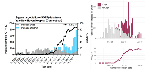
SGTF occurs when the TaqPath COVID-19 multiplex test has 2/3 targets successfully amplify when the S-gene target does not or “drops out.”  This phenomenon was first observed in the Alpha variant, because the probe for this target overlapped a characteristic mutation: S:Del69_70 (deletion of the 69th and 70th amino acids in the spike protein from a 6 base pair deletion). This mutation is absent in Delta, but present in Omicron, so has been used as an early tracker of Omicron prevalence.

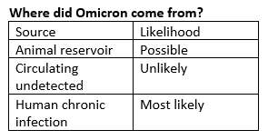
Most of this discussion is speculative and we won’t ever really know, but given the rate of transmission of this variant, it seems unlikely that it would have acquired so many mutations and not been detected before now. The most recent common ancestor is from over a year ago suggesting it was incubating for a long time.

We’ve seen a case of a person severely immunocompromised with no antibody response to vaccination + booster who still has an unmutated wild type strain in their system. With no immune pressure, the virus has not evolved.

However, in HIV+ patients with variable/ low immunity, there could be enough pressure to drive the immune evasion properties seen in Omicron. Southern Africa has over 30% of their HIV+ patients not on therapy who would be likely candidates for this type of host.

Did we see this coming?

Yes. Other immune evasive variants have arisen in areas with high prevalence of previous infection (Brazil/ S. Africa). Organisms evolve just enough to overcome the challenges in their environment. Thus the level of immunity provided by various immune exposures are approximately:

 Previous infection < 2x Vaccine < 2x Vaccine+ previous infection ~ x3 Vaccine

Scientists theorized that either Delta would evolve more immune evasive mutations or a totally new variant would arise. However, I didn’t think it would spread this quickly.

What is the impact?

Therapies. Most antibody therapies are directed as the business end of the spike protein—the receptor binding domain (RBD). The rest of the protein is covered in glycosylation modifications that block much recognition. Thus with many mutations in Omicron compared to the wild type strain (white), most therapeutic antibodies no longer bind/ inactivate viral replication.

Source: https://biorxiv.org/content/10.1101/2021.12.12.472269v1.full.pdf

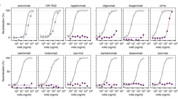
Only one monoclonal antibody—Sotrovimab from GSK—is effective, because it binds a pan-coronovirus epitope outside of the RBD. However, this antibody is in short supply.

  • Thus, knowing which variant someone has can direct therapy. Several hospitals in our area are out of Sotrovimab, and only people with the Delta variant can access other options. Thus, knowing the variant in a short time frame has clinical implications.
  • Whole genome sequencing takes too long, so the FDA has agreed to review PCR genotyping approaches for clinical use. I have described some previous approaches, but many of these methods are useful as a screening method and would not have sufficient specificity to determine whether an omicron variant is present. Next time, I will discuss variant genotyping, why it is important, how it can be done, and what clinical actions can be taken with the knowledge.

Severity. There are signs that it is less severe. Is this due to increase in immune tolerance? We now have been prepared by either previous infection or vaccination to be protected from hospitalization or severe disease.

@Jburnmurdoch https://twitter.com/jburnmurdoch/status/1478339769646166019/photo/1

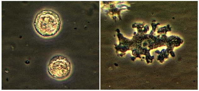
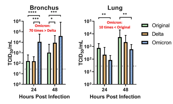
Or is the decline in severity due to lower pathogenicity? A recent non-peer reviewed study indicates the virus replicates x70 faster than Delta in the upper airways (left), but infiltrates cells 10% as well as the original strain.

From: https://www.med.hku.hk/en/news/press/20211215-omicron-sars-cov-2-infection?utm_medium=social&utm_source=twitter&utm_campaign=press_release

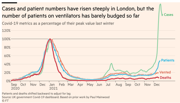
We all hope this will continue to be better news about the severity of Omicron, but from the lab side, I’ve heard of positivity rates >50% at some places. So this can still have a broad impact.

-Jeff SoRelle, MD is Assistant Professor of Pathology at the University of Texas Southwestern Medical Center in Dallas, TX working in the Next Generation Sequencing lab. His research interests include the genetics of allergy, COVID-19 variant sequencing, and lab medicine of transgender healthcare. Follow him on Twitter @Jeff_SoRelle.

Microbiology Case Study: A 36 Year Old Male Traveler with Fever

Case Description

A 36 year old male presented to the emergency department with complaints of fevers, chills, night sweats, nausea, diarrhea, weakness, and decreased appetite for 6 days. He often travels between India and Dallas, and five months prior to presentation returned from two years abroad. While overseas, he developed similar symptoms, but due to COVID-19 restrictions, he was unable to see a provider at that time. His family doctor prescribed a course of medication for presumed malaria, which he completed but could not recall the name of the medication. He endorsed being ill for two weeks at that time and improved with medication to complete resolution of his symptoms. Prior to presentation, he also endorsed 3-4 episodes of non-bloody diarrhea per day and remembered a period of self-resolving chills a month prior. His fever and rigors were cyclic, occurring every other day, worsening up to presentation.

Given his travel history and symptomology, blood was drawn in the emergency department for analysis including a malaria smear. CBC and CMP were significant for elevated bilirubin (Total bilirubin 1.6 mg/dL, Direct bilirubin 0.4 mg/dL), leukopenia (3.60 x 10(9)/L), macrocytosis (92.5 femtoliters), thrombocytopenia (86 x 10(9)/L), and elevated CRP (6.6 mg/dL). His blood differential was significant for neutrophilia (91%), lymphocytopenia (7%), and monocytopenia (1%). The malaria smear was positive, and the patient was given a dose of artemether/lumefantrine in the emergency department. Plasmodium vivax was identified at a parasitemia of 0.5% (Figure 1), and the infectious disease service recommended admission for further workup including testing for G6PD deficiency prior to starting primaquine. He was not G6PD deficient, and an ultrasound of the spleen was unremarkable. The patient was treated with chloroquine for the erythrocytic and primaquine for the exo-erythrocytic stages of P. vivax malaria.

Figure 1. Photomicrograph of Plasmodium vivax ring forms observed in this patient’s Giemsa-stained peripheral blood smear, which are counted to determine the level of parasitemia in a patient’s bloodstream (500x oil immersion).

Discussion

Malaria is an infection caused by protozoan parasites of the genus Plasmodium. These organisms are transmitted by female Anopheles mosquitos upon taking a blood-meal. Human malaria is caused by five defined Plasmodium species: P. falciparum, P. vivax, P. ovale, P. malariae, and P. knowlesi.1 While not endemic to the United States, there is significant disease burden worldwide. In 2019, an estimated 230 million cases of malaria were reported causing approximately 409,000 deaths.2

The two lifecycles of Plasmodium sp. in the human host are classically defined as “erythrocytic” and “exo-erythrocytic”, involving red blood cells and hepatocytes, respectively. Plasmodium sporozoites are inoculated into the human host from the salivary glands of the mosquito upon feeding. From there, the sporozoites travel to the liver where they infect hepatocytes, mature into schizonts and ultimately merozoites. The infected hepatocyte then ruptures, releasing merozoites which enter the circulation and infect erythrocytes, initiating the erythrocytic cycle. This is a unifying trait of all Plasmodium sp. causing human malaria. Importantly, P. vivax and P. ovale form hypnozoites (dormant forms) in the liver, which can reactivate (oftentimes months to years later) following bloodstream clearance, resulting in relapse. It is therefore important that Plasmodium sp. infections be accurately speciated, as management of liver stage parasites differs from that of those in the bloodstream. By contrast, P. malariae and P. falciparum do not form hypnozoites and thus do not chronically infect the liver.

Plasmodium speciationis accomplished by evaluating thin and thick blood spears,4 allowing for assessment of parasite morphology and determination of parasitemia to guide patient management. In cases of P. vivax, the red blood cells are often enlarged (1.5 to 2 times the size of uninfected erythrocytes). Ring forms in all stages of development can be observed in P. vivax infection. These ring forms subsequently mature into trophozoites or gametocytes. P. vivax trophozoites exhibit a large, amoeboid cytoplasm, large chromatin dots, and fine yellow-brown pigment. Trophozoites subsequently develop into schizonts in the infected erythrocytes, subsequently rupturing leading to autoinfection. P. vivax schizonts are large with coalesced pigment and harbor 12 or more merozoites3 (Figure 2). P. vivax gametocytes are large and round to oval shaped and have scattered brown pigment, hemozoin, that may fill the erythrocyte (Figure 3). Gametocytes will migrate to the capillaries which are taken up by a mosquito upon taking a blood-meal, completing the Plasmodium lifecycle.

Figure 2. Photomicrograph of Plasmodium vivax merozoites in a schizont (1000x oil immersion) from this patient.
Figure 3. Photomicrograph of Plasmodium vivax gametocyte with malaria pigment (500x oil immersion) from this patient.

Here we present a case of relapsed P. vivax infection. Blood stage P. vivax parasites are susceptible to chloroquine, but dormant hypnozoites in the liver are resistant to its effects. Hypnozoites can be treated with primaquine, and thus routine management of either P. ovale or P. vivax usually consists of a combination of both antimalarial drugs. It is important to note that primaquine is contraindicated in cases of G6PD deficiency and pregnancy due to hemolytic complications,2 which is why this patient was tested prior to initiating therapy.

P. vivax has a worldwide distribution but has higher prevalence in colder climates as compared to other malaria species. P. vivax is most commonly encountered in Latin America and Southeast Asia. In addition to colder climate adaptation, P. vivax is interesting in that the parasite uses Duffy red cell antigens to enter erythrocytes and in populations with low frequency of Duffy on the surface of RBCs those groups are generally resistant to P. vivax infection. However, there have been rare cases of P. vivax in Africans who are Duffy-null.5

References

  1. Gladwin, M., Mahan, C. S., & Trattler, B. (2021). Malaria. In Clinical microbiology made ridiculously simple (pp. 343–346). essay, MedMaster, Inc.
  2. Menkin-Smith L, Winders WT. Plasmodium Vivax Malaria. [Updated 2021 Jul 23]. In: StatPearls [Internet]. Treasure Island (FL): StatPearls Publishing; 2021 Jan-. Available from: https://www.ncbi.nlm.nih.gov/books/NBK538333/
  3. Procop, G. W., Koneman, E. W., & Winn, W. C. (2017). Malaria. In Koneman’s color Atlas and textbook of diagnostic microbiology (pp. 1467–1470). essay, Lippincott Williams & Wilkins.
  4. Laboratory diagnosis of malaria: Plasmodium vivax. Laboratory Identification of Parasites of Public Health Concern. (n.d.). Retrieved September 14, 2021, from https://www.cdc.gov/dpdx/resources/pdf/benchAids/malaria/Pvivax_benchaidV2.pdf.
  5. Gunalan, K., Niangaly, A., Thera, M. A., Doumbo, O. K., & Miller, L. H. (2018). Plasmodium vivax infections of duffy-negative erythrocytes: Historically undetected or a recent adaptation? Trends in Parasitology, 34(5), 420–429. https://doi.org/10.1016/j.pt.2018.02.006

-Elisa Lin is a fourth-year medical student at UT Southwestern Medical Center in Dallas, Texas. She is interested in AP/CP track residencies.

-Clare McCormick-Baw, MD, PhD is an Assistant Professor of Clinical Microbiology at UT Southwestern in Dallas, Texas. She has a passion for teaching about laboratory medicine in general and the best uses of the microbiology lab in particular.

-Andrew Clark, PhD, D(ABMM) is an Assistant Professor at UT Southwestern Medical Center in the Department of Pathology, and Associate Director of the Clements University Hospital microbiology laboratory. He completed a CPEP-accredited postdoctoral fellowship in Medical and Public Health Microbiology at National Institutes of Health, and is interested in antimicrobial susceptibility and anaerobe pathophysiology.

Microbiology Case Study: A Middle-Aged Man with Generalized Weakness

Case History

A middle age male with a past medical history of liver cirrhosis presented to the emergency department with one day of fever, chills, generalized weakness, and nausea. Complete blood count with differential showed leukopenia and neutropenia. Infectious work up was initiated including collection of 2 sets of blood cultures and imaging studies. A computed tomography (CT) scan of the abdomen revealed an irregularly shaped hypodense lesion in the right hepatic lobe concerning for abscess (Image 1). Ultrasound guided aspiration for the hepatic lesion yielded cloudy yellow bilious fluid, which was sent to the microbiology lab for aerobic and anaerobic cultures.

Image 1. CT scan of abdomen showing irregularly shaped hypodense lesion (yellow circle).

Two sets of blood cultures turned positive and Gram stain showed slender Gram positive rods in chains (Image 2). The aspirated fluid culture also showed 4+ Gram positive rods. Small gray colonies appeared on blood agar, chocolate agar, and Columbia Naladixic Acid (CNA) agar from both specimen types (Image 3). Lactobacillus rhamnosus was identified by matrix-assisted laser desorption/ionization time-of-flight mass spectrometry (MALDI-TOF MS). Minimal inhibitory concentration (MIC) was determined by broth microdilution assay and the organism was susceptible to penicillin while resistant to vancomycin. With appropriate antibiotics and abscess drainage, the patient’s condition improved and he was discharged to home.

Image 2. Gram stain of blood culture demonstrating gram positive rods in chains.
Image 3. Small gray colonies in blood agar from blood culture.

Discussion

Lactobacillus species are facultatively anaerobic, gram positive, non-spore forming rods that can have varying Gram stain morphology including short plump rods or long slender rods in chains or palisides.1 Lactobacillus species are bacterial inhabitant of the human mouth, gastrointestinal tract, and female genital tract. Isolation from clinical specimens could be considered by many to have questionable clinical significance.2 Lactobacillus species are often present in probiotics and fermented dairy products, including yogurt, and have been reported to provide benefits in gastrointestinal health,3–5 which led to increase in consumption of these products by many people, including our patient.

Cases of liver abscess and bacteremia caused by Lactobacillus species have been rarely reported in the literature and risk factors for the infection were immunosuppression, uncontrolled diabetes, hepatopancreaticobiliary disease, bacterial translocation, and use of probiotics or heavy dairy product consumption.6,7 The causative strains included L. rhamnosus, L. acidophilus, and L. paracasei.7

Pathophysiology of liver abscess and bacteremia due to Lactobacillus species is not well understood but it is postulated that several mechanisms may contribute to the pathogenicity of lactobacilli. Some strains are able to bind to intestinal mucosa, which may aid in translocation of the organism into the bloodstream. Also some strains can adhere to extracellular matrix proteins, aggregate platelets, and produce glycosidases and proteases.7 Furthermore, some strains are more resistant to intracellular killing by macrophages and nitric oxide.8

It is worth noting that many species of Lactobacillus are intrinsically resistant to vancomycin. However, they are usually susceptible to penicillin and ampicillin, as it was seen in our patient, and antimicrobial susceptibility testing can be performed by determining MIC of antimicrobials.9

References

  1. Goldstein EJC, Tyrrell KL, Citron DM. Lactobacillus Species: Taxonomic Complexity and Controversial Susceptibilities. Clin Infect Dis. 2015;60(suppl_2):S98-S107. doi:10.1093/CID/CIV072
  2. Chan JFW, Lau SKP, Woo PCY, et al. Lactobacillus rhamnosus hepatic abscess associated with Mirizzi syndrome: a case report and review of the literature. Diagn Microbiol Infect Dis. 2010;66(1):94-97. doi:10.1016/J.DIAGMICROBIO.2009.08.009
  3. Kligler B, Cohrssen A. Probiotics. Am Fam Physician. 2008;78(9):1073-1078. Accessed November 17, 2021. http://www.aafp.org/afp.
  4. Anukam KC, Osazuwa EO, Osadolor HB, Bruce AW, Reid G. Yogurt containing probiotic Lactobacillus rhamnosus GR-1 and L. reuteri RC-14 helps resolve moderate diarrhea and increases CD4 count in HIV/AIDS patients. J Clin Gastroenterol. 2008;42(3):239-243. doi:10.1097/MCG.0B013E31802C7465
  5. Adolfsson O, Meydani SN, Russell RM. Yogurt and gut function. Am J Clin Nutr. 2004;80(2):245-256. doi:10.1093/AJCN/80.2.245
  6. Omar AM, Ahmadi N, Ombada M, et al. Breaking Bad: a case of Lactobacillus bacteremia and liver abscess. J Community Hosp Intern Med Perspect. 2019;9(3):235. doi:10.1080/20009666.2019.1607704
  7. Sherid M, Samo S, Sulaiman S, Husein H, Sifuentes H, Sridhar S. Liver abscess and bacteremia caused by lactobacillus: Role of probiotics? Case report and review of the literature. BMC Gastroenterol. 2016;16(1):1-6. doi:10.1186/S12876-016-0552-Y/TABLES/1
  8. Asahara T, Takahashi M, Nomoto K, et al. Assessment of Safety of Lactobacillus Strains Based on Resistance to Host Innate Defense Mechanisms. Clin Diagn Lab Immunol. 2003;10(1):169. doi:10.1128/CDLI.10.1.169-173.2003
  9. CLSI. M45. Methods for Antimicrobial Dilution and Disk Susceptibility Testing of Infrequently Isolated or Fastidious Bacteria ; Proposed Guideline. Vol 35.; 2015. Accessed November 17, 2021. http://www.clsi.org.

Do Young Kim, MD is a medical microbiology fellow at University of Chicago (NorthShore). His academic interests include clinical microbiology and infectious diseases, epidemiology, and public health.

-Paige M.K. Larkin, PhD, D(ABMM), M(ASCP)CM is the Director of Molecular Microbiology and Associate Director of Clinical Microbiology at NorthShore University HealthSystem in Evanston, IL. Her interests include mycology, mycobacteriology, point-of-care testing, and molecular diagnostics, especially next generation sequencing.

Microbiology Case Study: Not An Ordinary Sore Throat, but One Accompanied by Headache 

Case History

An 18 year old healthy female presented to the emergency department of a tertiary care hospital in Minnesota for headache, vomiting, and sore throat. She did not have any significant past medical history. Due to meningitis concerns, lumbar puncture and head computed tomography (CT) imaging were performed. The CT scan showed an accumulation of fluid in the posterior right frontal sinus with scattered mucosal thickening. However, her cerebrospinal fluid (CSF) profile was insignificant, with normal protein and glucose levels. CSF culture was ordered, and two sets of blood cultures were drawn. 

Based on the examination and presenting symptoms, pharyngitis was suspected, and she was discharged with Amoxicillin (500mg Q6H for five days). However, her strep throat screening returned negative. Her blood culture was negative. CSF culture was also negative. Cryptococcal antigen and Enterovirus PCR were performed; however, both results were negative.

She returned to the ED two days later for a worsening headache and newly developed photophobia. Additional history revealed that she went swimming in a lake two weeks prior to her first presentation at the ED. Her CSF was sent for the Ova and Parasite (O&P) exam for suspicious parasitic meningitis. The CSF O&P test was negative. CSF PCR for amoeba was also performed at a reference laboratory, and the results came back positive with Balamuthia mandrillaris. The patient was then given flucytosine, fluconazole, and azithromycin. 

Figure 1. Photo Credit CDC: Balamuthia – free living parasite observed under light microscopy.

Discussion

Balamuthia mandrillaris belongs to a group of free-living amoebae, including Acanthamoeba species and Naegleriafowleri, that cause fatal encephalitis.1 Balamuthia mandrillaris is the only known species of the genus Balamuthia that causes infections in humans. Encephalitis caused by B. mandrillaris is known as granulomatous amoebic encephalitis (GAE). GAE is characterized as a subacute to a chronic infection that can last several months to years.2 GAE differs from primary amoebic meningoencephalitis (PAM) caused by Naegleria fowleri, which typically causes an acute onset lasting a few days. 

While ecological niches of B. mandrillaris are not well understood, they have been reported to be isolated from dust, soil, and water.r Both trophozoite and cyst forms can enter the body through the nasal passage or ulcerated/broken skin; however, the trophozoite stage causes associated disease manifestation and represents a diagnostic stage.2 

Brain-eating amoebas are traditionally difficult to diagnose. Hematology and chemistry profiles of CSF of affected individuals are generally unremarkable, although, sometimes, increased monocytes and lymphocytes, along with increased protein levels, are seen in some cases of GAE.3 

The most common method of laboratory diagnosis of B. mandrillaris is a microscopic examination of CSF wet mount (Figure 1) or via immunohistochemical staining of CSF or brain biopsy.1 With advancements in technology, species-specific nucleic acid amplification tests (NAAT) can be performed to diagnose B. mandrillaris infection accurately. However, there is no commercially available NAAT for the free-living amoeba. Only very few laboratories, such as State departments of health laboratories, Centers for Disease Control (CDC) and Prevention, or commercial reference laboratories, develop these tests as a laboratory-developed test (LDT). Histological assessment of biopsies from brain lesions may reveal tumor-like appearance or perivascular monocytic necrosis of affected areas.1 While there have been significant technological advancements, the prognosis stays at less than a 5% survival rate,6 with only roughly 25% of cases diagnosed antemortem. One possible reason for delayed laboratory diagnosis is the challenges in performing the microscopic examination in clinical microbiology laboratories since it requires expertise for accurate identification of the organism. Additionally, most clinical microbiology laboratories do not readily have an in-house LDT for free-living amoeba NAAT. Therefore, the turnaround time for diagnosing B. mandrillaris or any free-living amoeba is typically longer when specimens have to be sent out to reference laboratories. 

Diagnosis of B. mandrillaris encephalitis solely based on clinical symptoms is often challenging due to similar presentation in other causes of infectious encephalitis. B. mandrillaris can affect both immunocompetent and immunocompromised individuals.1,3,4 The first B. mandrillaris case was reported in a deceased baboon in the San Diego Zoo in 1986.1  The majority of patients were diagnosed postmortem.1 While most B. mandrillaris infections are actively acquired through nasal passages or skin penetration, rare post-mortem cases of passive transfer of the organism from organ transplantation have been reported.6 With technological advancement, there have been successes in pre-mortem diagnoses in recent years.1,4 According to the known cases, individuals of Latin American origin are more likely to contract the disease; it is unknown if it is due to increased exposure or a genetic predisposition.1  Similar to other free-living amoebae, B. mandrillaris can be generally found in warmer climates or tropical regions. Of approximately two hundred cases reported worldwide, about 34 were reported in Latin America, from Mexico to Brazil, while some were from Japan, New Zealand, England, and other European countries. The Southwestern United States also contributes 30 cases, mostly in Arizona, Texas, and California.1  In the United States, there have only been 109 cases directly reported to CDC from 1974 to 2016.2,7  We believe that this is the first case of Balamuthia reported in Minnesota. The number of exact cases would be difficult to be determined due to misdiagnosis and rare occurrence of the disease or cases not reported to CDC or the state department of health. 

While investigational drugs for B. mandrillaris GAE are in development, combination therapy of flucytosine, fluconazole, pentamidine, and azithromycin or clarithromycin has shown successes.2 Our patient was successfully treated with flucytosine, fluconazole, and azithromycin. 

References

  1. Matin A, Siddiqui R, Jayasekera S, Khan NA. Increasing importance of Balamuthia mandrillaris. Clin Microbiol Rev. 2008 Jul;21(3):435-48. doi: 10.1128/CMR.00056-07. PMID: 18625680; PMCID: PMC2493082. 
  2. Centers for Disease Control and Prevention. (2019, August 23). CDC – Dpdx – free Living Amebic Infections. Centers for Disease Control and Prevention. https://www.cdc.gov/dpdx/freelivingamebic/index.html.
  3. Kofman A, Guarner J. Free Living Amoebic Infections: Review. J Clin Microbiol. 2021 Jun 16:JCM0022821. doi: 10.1128/JCM.00228-21. Epub ahead of print. PMID: 34133896.
  4. Pietrucha-Dilanchian, P., Chan, J. C., Castellano-Sanchez, A., Hirzel, A., Laowansiri, P., Tuda, C., Visvesvara, G. S., Qvarnstrom, Y., & Ratzan, K. R. (2011). Balamuthia mandrillaris And Acanthamoeba Amebic Encephalitis With Neurotoxoplasmosis Coinfection in a patient with Advanced HIV Infection. Journal of Clinical Microbiology, 50(3), 1128–1131.
  5. Ong TYY, Khan NA, Siddiqui R. 2017. Brain-eating amoebae: predilection sites in the brain and disease outcome. J Clin Microbiol 55:1989 –1997. https://doi.org/10.1128/JCM. 02300-16.
  6. Centers for Disease Control and Prevention. 2011. Balamuthia mandrillaris transmitted through organ transplantation—Mississippi, 2009. Am J Trans-plant 11:173–176. https://doi.org/10.1111/j.1600-6143.2010.03395_1.x.
  7. Jennifer R Cope, Janet Landa, Hannah Nethercut, Sarah A Collier, Carol Glaser, Melanie Moser, Raghuveer Puttagunta, Jonathan S Yoder, Ibne K Ali, Sharon L Roy, The Epidemiology and Clinical Features of Balamuthia mandrillaris Disease in the United States, 1974–2016, Clinical Infectious Diseases, Volume 68, Issue 11, 1 June 2019, Pages 1815–1822, https://doi.org/10.1093/cid/ciy813

-Alejandro Soto, MLS (ASCP)CM is a junior medical technologist who is passionate about clinical microbiology.

-Phyu M. Thwe, Ph.D., D(ABMM), MLS(ASCP)CM is Microbiology Technical Director at Allina Health Laboratory in Minneapolis, MN. She completed her CPEP microbiology fellowship at the University of Texas Medical Branch in Galveston, TX. Her interest includes appropriate test utilization and extra-pulmonary tuberculosis.