A 15 month patient was seen in the Pediatric Hematology-Oncology clinic in June 2014 for mild normocytic anemia.
Review of Systems
Negative 12 system review. No history of pallor, jaundice or high colored urine.
| Ref. Range | 6/14 | |
| WBC | 6-17 K/uL | 11.4 |
| Hemoglobin | 10.5-13.5 g/dL | 8.9 (L) |
| Hematocrit | 33-39 % | 25.4 (L) |
| Platelets | 150-400 K/uL | 648 (H) |
| RBC | 3.7-5.3 M/uL | 3.57 (L) |
| MCV | 70-86 fL | 71.2 |
| MCH | 23-30 pg | 24.9 |
| MCHC | 31-36 % | 34.9 |
| RDW | 11.5-14.5 % | 16.4 (H) |
His serum iron profile was normal, serum lead levels were normal. Reticulocyte percentage and absolute reticulocyte count were also both not elevated.
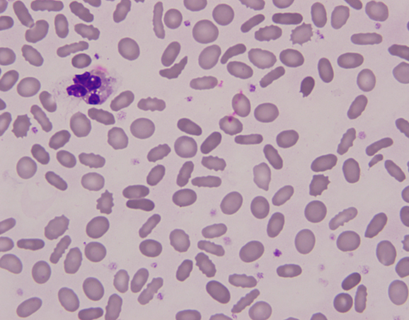
Review of peripheral smear revealed moderate anisopoikilocytosis with presence of numerous elliptocytes.

Molecular studies demonstrated a heterozygous mutation in the EPB41 gene associated with HE.
Patient was diagnosed with Hereditary Elliptocytosis (HE).
He has been followed up at the hematology clinic for a year now. His follow up CBC results are as follows. He has reached his age appropriate milestones and continues to grow well.
| Ref. Range | 7/14 | 10/14 | 12/14 | 4/15 | |
| WBC | 6-17 K/uL | 10.3 | 11.3 | 7.4 | 10.0 |
| Hemoglobin | 10.5-13.5 g/dL | 9.3 (L) | 9.9 (L) | 9.7 (L) | 11.0 |
| Hematocrit | 33-39 % | 26.5 (L) | 28.7 (L) | 28.1 (L) | 33.6 |
| Platelets | 150-400 K/uL | 599 (H) | 570 (H) | 403 (H) | 447 (H) |
| RBC | 3.7-5.3 M/uL | 3.74 | 3.91 | 3.87 | 4.58 |
| MCV | 70-86 fL | 70.8 | 73.3 | 72.6 | 73.5 |
| MCH | 23-30 pg | 24.8 | 25.4 | 25.1 | 23.9 |
| MCHC | 31-36 % | 35.0 | 34.7 | 34.5 | 32.6 |
| RDW | 11.5-14.5 % | 16.9 (H) | 17.7 (H) | 18.2 (H) | 18.0 (H) |
Hereditary elliptocytosis (HE) is an inherited hemolytic anemia, secondary to red cell membrane defect more commonly assembly of spectrin, spectrin-ankyrin binding, protein 4.1 and glycophorin C with a clinical severity ranging from asymptomatic carriers to a severe hemolytic anemia. It is more common in individuals from African and Mediterranean decent – neither applies to our patient.It is inherited in an autosomal dominant pattern, typically individual who are heterozygous are asymptomatic while those who are homozygous or compound heterozygous have a mild to severe anemia. Occasional patients with more severe hemolysis may require splenectomy.
Regardless of the underlying molecular abnormality, most circulating red cells are elliptical or oval. They still have an area of central pallor, since there is no loss of the lipid bilayer (as seen in Hereditary spherocytosis).

-Neerja Vajpayee, MD, is an Associate Professor of Pathology at the SUNY Upstate Medical University, Syracuse, NY. She enjoys teaching hematology to residents, fellows and laboratory technologists.
