In several previous blogs, I’ve mentioned the topic of post-mortem radiography (or “x-rays”). While postmortem CT scanning is a hot topic in the field, plain films are a tool which has been in widespread use for decades. Autopsy standards of the National Association of Medical Examiners require, at a minimum, radiographs be performed on all infants, gunshot wound victims, explosion victims, and charred or decomposed remains. Let’s examine the reasons for these requirements and look at a few specific examples.
All infants must get full body x-rays to check for acute or healing rib and long bone (extremity) fractures, which could be indicative of physical abuse. The extremities are not usually dissected in the course of a typical post-mortem examination, but fractures that can’t be attributed to birth trauma (especially in a pre-mobile child) are a concerning finding that needs further investigation and dissection.
Radiographs taken of gunshot wound victims document the presence and location of retained projectiles, all of which need to be recovered as evidence.

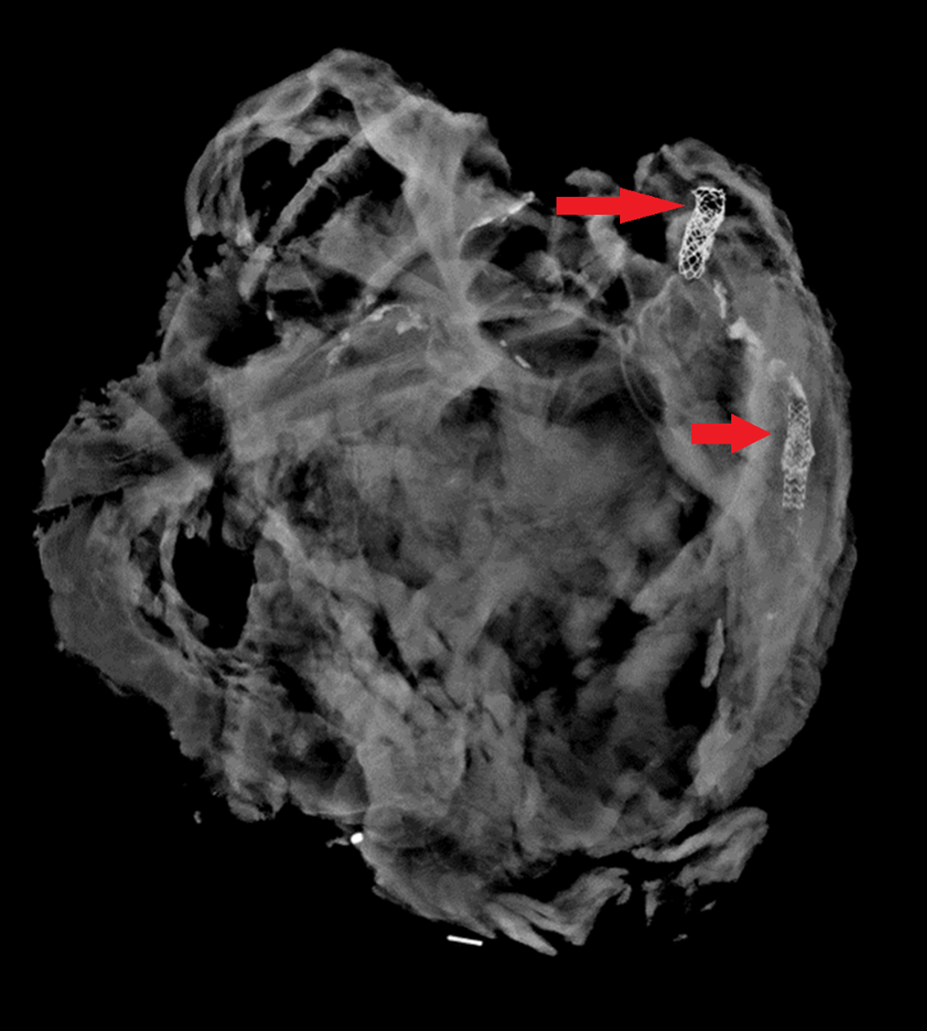
Radiographs are commonly performed in any type of penetrating trauma, including sharp force injury and explosive injuries, to identify retained foreign bodies (especially broken fragments of the blade, which may pose a risk to the pathologist). Similar to projectiles, these fragments need to be recovered as evidence. Radiographs can also document the presence of an air embolism, which can be missed at autopsy if special dissection techniques aren’t performed.
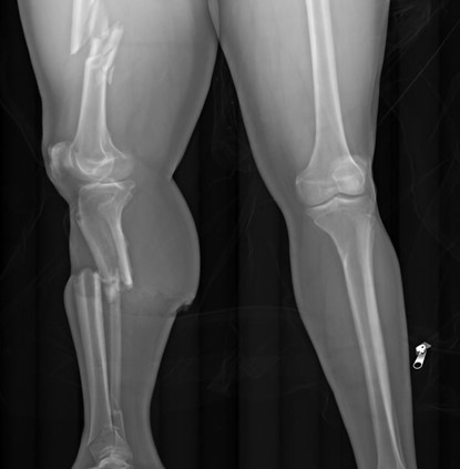
In pedestrians who have been struck by motor vehicles, radiography is the first step in examining trauma. As mentioned earlier, the extremities aren’t typically dissected during a traditional autopsy – but in pedestrians, lower extremity fractures can document the site of initial impact, and the distance of the fracture from the foot may indicate the bumper height of the car.
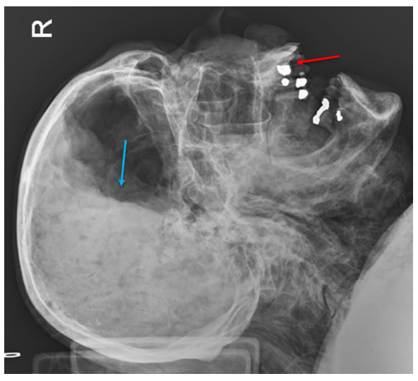
Fire victims may have extensive thermal injuries and charring which can hide evidence of other injuries, and radiographs can help identify them. Radiographs are also one step toward identifying the victim. Decomposed bodies must be x-rayed for similar reasons – external and internal soft tissue alterations make assessment for trauma more difficult, and radiographs may be needed to confidently identify the body.
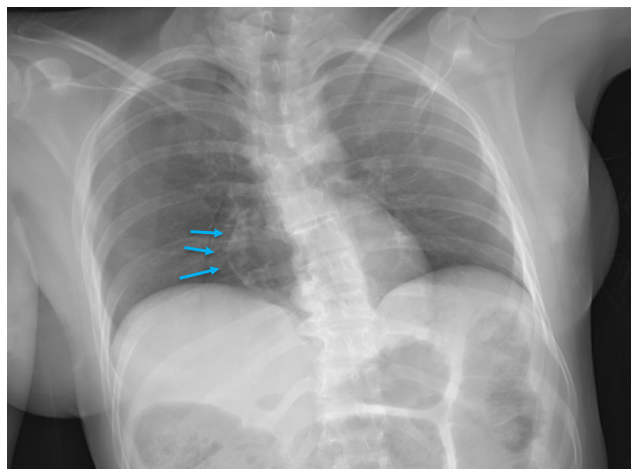
On occasion, radiographs can be performed on individual organs to help define anatomy or previous surgical alterations. Especially in the heart, radiographs can demonstrate coronary artery or valve calcifications, surgical clips from bypass grafts, or other radiopaque prostheses. Knowing the location of devices before dissection gives the pathologist a better chance at preserving and evaluating important structures.
Not all offices can afford the installation (or maintenance) of CT scanners, but access to x-ray machines is more widespread. As we’ve seen here, x-rays are a versatile tool which can document injuries, help identify decedents, and direct the pathologist to perform special autopsy procedures which aren’t part of the daily routine.

-Alison Krywanczyk, MD, FASCP, is currently a Deputy Medical Examiner at the Cuyahoga County Medical Examiner’s Office.
