Microbiology Case Study: A Middle-Aged Male with Altered Mental Status

Case presentation

A middle-aged male with a history of type 2 diabetes, hypertension, obesity, and alcohol-related cirrhosis presented to the emergency department with altered mental status. He was obtunded, acutely encephalopathic and hypoglycemic. He soon developed emesis and coded during clinical assessment, undergoing emergent intubation. He was found to be profoundly acidotic with labs consistent with disseminated intravascular coagulation and multiorgan failure. The patient was transfused but continued to code multiple times before and during ICU transfer/admission. Despite multiple resuscitation attempts, he expired soon afterwords.

Laboratory workup

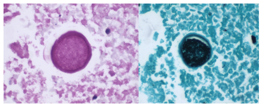
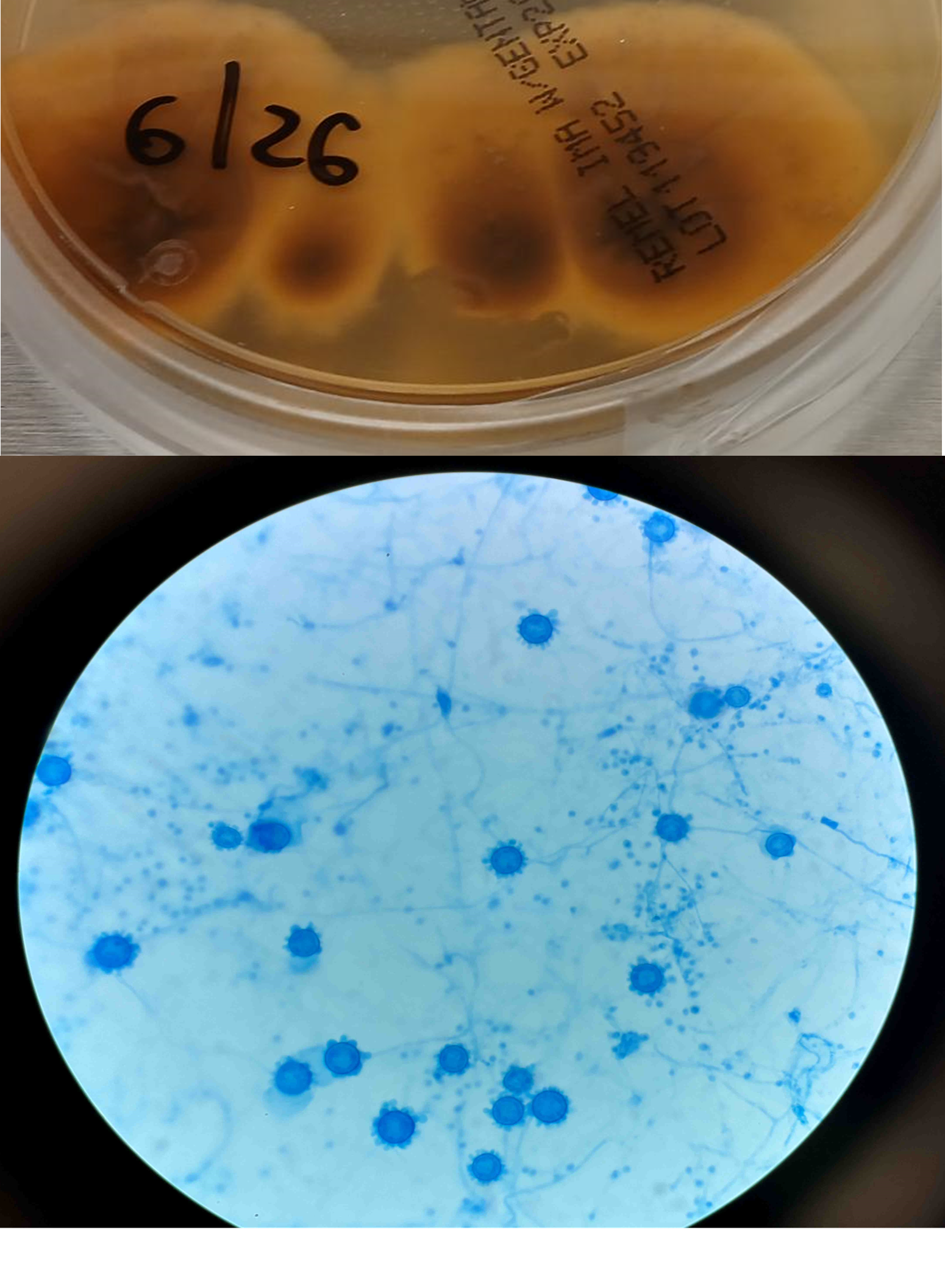
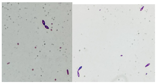
Blood cultures drawn in the ED prior to admission became positive with curved gram-negative rods (Image 1A) within 16 hours. An oxidase-positive, indole-positive, beta-hemolytic organism was recovered after 24 hours of incubation. The organism was lactose-fermenting (confirmed by ONPG) and exhibited colorless growth on Thiosulfate Citrate Bile Salts agar suggestive of a lack of sucrose utilization (Image 1B, set up for demonstrative purposes). The organism was definitively identified as Vibrio vulnificus by MALDI-TOF MS. No additional workup was undertaken as the patient had expired prior to the organism being recovered from blood culture.

Image 1.  A: Gram stain from a positive blood bottle revealing curve gram-negative rods (100X magnification). Inset demonstrates the morphologically distinct curved appearance.  B: Blood and TCBS agars revealing growth of V. vulnificus. Lack of yellow colorization on TCBS media indicates lack of sucrose utilization. Biochemical testing revealed oxidase, catalase, and indole positivity also consistent with the MALDI-TOF identification of V. vulnificius.

Discussion

Vibrio sp. are marine bacteria that naturally colonize brackish and saltwater aquatic environments. Of the more than 70 currently recognized species, at least 12 are recognized as human pathogens.1 Human infections are broadly classified as being either cholera (caused by V. cholerae) or vibriosis (caused by other non-V. cholerae Vibrio spp.). Unlike cholera; a severe diarrheal illness usually acquired through ingestion of contaminated food or water, vibriosis represents a group of infections with varied clinical manifestations dependent upon the etiologic agent, route of infection, and host susceptibility.2 Non-cholera vibrios are often found in seawater with moderate to high salinity and as clinically important contaminants of raw or undercooked seafood.

                V. vulnificus thrives in warmer water and infections follow a seasonality, peaking in the warmer summer months.3 In contrast to V. cholerae and V. parahaemolyticus, V. vulnificus infections generally are associated with patients with underlying conditions, most commonly diabetes, liver disease and iron storage disorders. It is estimated that patients with chronic liver diseases (particularly cirrhosis due to either alcoholism or chronic hepatitis B or C) are 80-fold more likely to develop V. vulnificus-associated primary septicemia than healthy counterparts.2 Infections are most common in men aged 45-60 years who make up 85-90% of patients,4 consistent with this case. Contaminated food consumption (particularly filter feeding shellfish) can result in gastroenteritis or primary septicemia and disseminated disease.

Non-cholera vibrios are estimated to cause up to 80,000 infections worldwide, with V. parahaemolyicus and V. alginolyticus responsible for most cases. Among this group of organisms, V. vulnificus stands out as being particularly virulent; between 150-200 V. vulnificus infections are reported to the US CDC annually, with 20% being fatal.5 This organism oftencauses more than 95% of seafood-related deaths in the United States and the highest case fatality of any foodborne pathogen [2]. V. vulnificus also causes serious skin/soft tissue infections. This can occur either through exposure of a preexisting wound to contaminated seawater, or through injury while handling contaminated seafood. Cutaneous infections can present as cellulitis or bullae, which can progress to necrotizing disease and secondary sepsis is left untreated.6

No epidemiological link was able to be established between this patient’s case and either seafood or exposure to seawater, although cases of V. vulnificus infections among patients without classical exposure risk has been documented.7 This case of V. vulnificus primary septicemia highlights the acuity of this presentation as well as the importance of including V. vulnificus in differential diagnosis of hosts with associated risk factors and/or epidemiological links. Blood culture remains the gold standard for diagnosis. Importantly, doxycycline in combination with a third-generation cephalosporin constitutes the standard regimen for antibiotic therapy – however, doxycycline is not usually considered a first-line antibiotic for management of patient with gram-negative bloodstream infections, so rapid and accurate identification of V. vulnificus in this setting is essential.

1.           Kokashvili, T., et al., Occurrence and Diversity of Clinically Important Vibrio Species in the Aquatic Environment of Georgia. Front Public Health, 2015. 3: p. 232.

2.           Baker-Austin, C., et al., Vibrio spp. infections. Nature Reviews Disease Primers, 2018. 4(1): p. 1-19.

3.           Hughes, M.J., et al., Notes from the Field: Severe Vibrio vulnificus Infections During Heat Waves – Three Eastern U.S. States, July-August 2023. MMWR Morb Mortal Wkly Rep, 2024. 73(4): p. 84-85.

4.           Jones, M.K. and J.D. Oliver, Vibrio vulnificus: disease and pathogenesis. Infect Immun, 2009. 77(5): p. 1723-33.

5.           Bharathan, A., et al., Implication of environmental factors on the pathogenicity of Vibrio vulnificus: Insights into gene activation and disease outbreak. Microb Pathog, 2025. 204: p. 107591.

6.           Coerdt, K.M. and A. Khachemoune, Vibrio vulnificus: Review of Mild to Life-threatening Skin Infections. Cutis, 2021. 107(2): p. E12-e17.

7.           Candelli, M., et al., Vibrio vulnificus—A Review with a Special Focus on Sepsis. Microorganisms, 2025. 13(1): p. 128.

-Rene Bulnes, MD is an Infectious Diseases Clinician and current Medical Microbiology Fellow at the University of Texas Southwestern Medical enter in Dallas, TX.

-Andrew Clark, PhD, D(ABMM) is an Assistant Professor at the Johns Hopkins University School of Medicine in the Department of Pathology, and Director of the Bacteriology Laboratory at the Johns Hopkins Hospital. He completed a CPEP-accredited postdoctoral fellowship in Medical and Public Health Microbiology at National Institutes of Health, and is interested in the molecular mechanisms of antimicrobial resistance, susceptibility testing, and the evaluation of novel technology for the clinical microbiology laboratory.

Microbiology Case Study: Diarrhea in a Patient with Renal Transplant

Case History

            A-58-year-old female with past medical history of breast cancer in remission, renal transplant 8 years ago on tacrolimus, now presenting with inability to tolerate oral intake, dyspnea on exertion and gastrointestinal symptoms such as profuse foul-smelling, non-bloody diarrhea, and vomiting. She denied exposure to sick contacts, recent travel, or changes in diet. She had other co-morbidities including hypertension, atrial arrhythmia, hyperlipidemia, and past C. difficile infection. On the physical exam, she was afebrile, normotensive, with a normal heart rate and rhythm, and her abdomen is soft and non-distended, with tenderness to palpation. Cardiology was consulted due to new onset paroxysmal atrial fibrillation on telemetry, and the renal transplant team was contacted for admission in concern for her rising creatinine. Upon admission to the floor, the patient had 2 more loose stools which were collected for stool culture and multiplex, syndromic gastrointestinal PCR panel, which tested positive for norovirus. She was kept on contact precautions.

Discussion

            Norovirus is a single stranded positive-sense RNA virus belonging to the Calciciviridae family. Noroviruses are divided into 10 genogroups based on the amino acid sequence of VP1, the norovirus capsid protein. Genogroups GI and GII account for 90% of reported infections including outbreaks, with the GII.4 genotype being the cause of most severe disease, and it is more frequently implicated in outbreaks than other genotypes (1).

            The median incubation period for norovirus infections is 1.2 days, with most symptomatic patients presenting with diarrhea and vomiting. Asymptomatic shedding of norovirus is common, mainly in the pediatric population, where 11.6 – 49.2% of stool samples were found to contain norovirus in random recruitment studies globally. Most patients with norovirus infection have spontaneous resolution of symptoms by the third day of illness. Elderly, young, and immunocompromised patients face greater risk of severe and prolonged symptoms from norovirus infection. Chronic infection with norovirus may occur in immunocompromised hosts, with associated symptoms lasting up to weeks or months in these patients (2).

            Transmission of norovirus occurs through oral-oral and fecal-oral routes, and transmission can occur directly via exposure to human emesis or feces, or through contamination of food, water, or fomites with such samples. Median viral titers have been recorded at 3.9 x 10⁴ copies/mL in emesis samples from patients with norovirus GII, with aerosolization of viral particles possible due to projectile vomiting and toilet flushing (de Graaf). Once inoculated, norovirus particles primarily infect macrophages and dendritic cells in the gastrointestinal tract, with current reports suggesting that particles enter these cells via attachment to blood group antigens, or HGBAs, and Toll-like receptors (3). VP1, the capsid protein of norovirus, has been found to induce expression of aquaporin-1 in human intestinal cell culture. This leads to small molecule permeability at the intestinal barrier, which likely leads to the watery diarrhea seen in norovirus infection (4).

            Norovirus is implicated as the cause of 19% of all acute gastroenteritis cases globally, with virtually every country reporting norovirus cases (5). These infections frequently occur in the form of outbreaks, with the highest rates of infection recorded in the winter months (Figure 1). Crowded and closed environments, including daycare centers, cruise ships, and restaurants, are known facilitators of outbreaks. More than half of all norovirus outbreaks are reported in healthcare settings, such as hospitals and long-term care facilities (6).  

Figure 1. Increase in norovirus outbreaks was reported in 2025. (Data adapted from CDC: Norovirus Outbreaks Reported by State Health Departments. https://www.cdc.gov/norovirus/php/reporting/norostat-data-table.html)

Immunological assays (e.g., antigen detection) and transmission electron microscopy can be used for detection of gastrointestinal viruses, but these methods have limited sensitivity and specificity and not recommended for clinical diagnosis (7). Laboratory detection by molecular methods is the preferred method for norovirus diagnosis. Testing of stool specimens may be performed on single plex or FDA-approved, commercially available, multiplex syndromic PCR panels. While PCR is the most sensitive approach, false-positives have been reported (8). Five regions of the genome (A,B,C,D, and E) of the norovirus genome have been used for genotyping while viral capsid gene (encoded by regions C, D, and E) is typically used given the viral capsid being involved in host-receptor interactions and immune response (9). In certain instances, sequencing and detection of the ORF1 and ORF2 genes may help identify strains after antigenic drift events (10).

            Oral or intravenous rehydration is the mainstay of norovirus treatment in all patients. In immunocompetent patients, norovirus is expected to resolve spontaneously (11). Chronic symptomatic infection with norovirus in immunocompromised patients poses a significant clinical challenge, especially when reduction in immunosuppressants is not feasible. In such patients, case reports have suggested that nitazoxanide may achieve resolution of symptoms, and one retrospective study proposed that addition of metronidazole led to resolution of norovirus symptoms in nitazoxanide-refractory cases (12-14). However, no randomized controlled trials have demonstrated the efficacy of nitazoxanide or metronidazole in chronic norovirus infection. Norovirus vaccines are currently in development, but challenges include high genetic diversity of circulating strains, lack of understanding regarding herd immunity and correlates of immune response, and the lack of standardized testing approaches such as cell cultures or animal models for efficacy studies.

References

1. Carlson KB, Dilley A, O’Grady T, Johnson JA, Lopman B, Viscidi E. A narrative review of norovirus epidemiology, biology, and challenges to vaccine development. NPJ Vaccines. 2024;9(1):94. Published 2024 May 29. doi:10.1038/s41541-024-00884-2

2. Robilotti E, Deresinski S, Pinsky BA. Norovirus. Clin Microbiol Rev. 2015;28(1):134-164. doi:10.1128/CMR.00075-14

3. Chen J, Cheng Z, Chen J, Qian L, Wang H, Liu Y. Advances in human norovirus research: Vaccines, genotype distribution and antiviral strategies. Virus Res. 2024;350:199486. doi:10.1016/j.virusres.2024.199486

4. Zhang M, Zhang B, Chen R, et al. Human Norovirus Induces Aquaporin 1 Production by Activating NF-κB Signaling Pathway. Viruses. 2022;14(4):842. Published 2022 Apr 18. doi:10.3390/v14040842

5. Zhang P, Hao C, Di X, et al. Global prevalence of norovirus gastroenteritis after emergence of the GII.4 Sydney 2012 variant: a systematic review and meta-analysis. Front Public Health. 2024;12:1373322. Published 2024 Jun 27. doi:10.3389/fpubh.2024.1373322

6. Tsai H, Yune P, Rao M. Norovirus disease among older adults. Ther Adv Infect Dis. 2022;9:20499361221136760. Published 2022 Nov 14. doi:10.1177/20499361221136760

7. Rabenau HF, Stürmer M, Buxbaum S, Walczok A, Preiser W, Doerr HW. Laboratory diagnosis of norovirus: which method is the best? Intervirology. 2003;46(4):232-8. doi: 10.1159/000072433.

8. Caza M, Kuchinski K, Locher K, Gubbay J, Harms M, Goldfarb DM, Floyd R, Kenmuir E, Kalhor M, Charles M, Prystajecky N, Wilmer A. Investigation of suspected false positive norovirus results on a syndromic gastrointestinal multiplex molecular panel. J Clin Virol. 2024 Dec;175:105732. doi: 10.1016/j.jcv.2024.105732. Epub 2024 Sep 30. 

9. Mattison K, Grudeski E, Auk B, Brassard J, Charest H, Dust K, Gubbay J, Hatchette TF, Houde A, Jean J, Jones T, Lee BE, Mamiya H, McDonald R, Mykytczuk O, Pang X, Petrich A, Plante D, Ritchie G, Wong J, Booth TF. Analytical performance of norovirus real-time RT-PCR detection protocols in Canadian laboratories. J Clin Virol. 2011 Feb;50(2):109-13. doi: 10.1016/j.jcv.2010.10.008. Epub 2010 Nov 10.

10. Mattison K, Grudeski E, Auk B, Charest H, Drews SJ, Fritzinger A, Gregoricus N, Hayward S, Houde A, Lee BE, Pang XL, Wong J, Booth TF, Vinjé J. Multicenter comparison of two norovirus ORF2-based genotyping protocols. J Clin Microbiol. 2009 Dec;47(12):3927-32. doi: 10.1128/JCM.00497-09. Epub 2009 Oct 21.

11. Mirza S, Hall A. Norovirus | CDC Yellow Book 2024. wwwnc.cdc.gov. Published May 1, 2023. https://wwwnc.cdc.gov/travel/yellowbook/2024/infections-diseases/norovirus

12. Haubrich K, Gantt S, Blydt-Hansen T. Successful treatment of chronic norovirus gastroenteritis with nitazoxanide in a pediatric kidney transplant recipient. Pediatr Transplant. 2018;22(4):e13186. doi:10.1111/petr.13186

13. Siddiq DM, Koo HL, Adachi JA, Viola GM. Norovirus gastroenteritis successfully treated with nitazoxanide. J Infect. 2011;63(5):394-397. doi:10.1016/j.jinf.2011.08.002

14. Soneji M, Newman AM, Toia J, Muller WJ. Metronidazole for treatment of norovirus in pediatric transplant recipients. Pediatr Transplant. 2022;26(8):e14390. doi:10.1111/petr.14390

-Brendan Sweeney is a third-year medical student at the George Washington University School of Medicine and Health Sciences. His research interests include infectious diseases, hematopathology, and point of care diagnostics.

-Rebecca Yee, PhD, D(ABMM), M(ASCP)CM is the Chief of Microbiology, Director of Clinical Microbiology and Molecular Microbiology Laboratory at the George Washington University Hospital. Her interests include bacteriology, antimicrobial resistance, and development of infectious disease diagnostics.

Microbiology case study: Not so-cording MTB in a case of pediatric TB meningitis

Case History

An 11-year-old girl was brought by her mother to the Emergency Room because of altered mental status, described as abnormal movements and staring with a period unresponsiveness lasting 15 minutes.

One week previously, she had returned from a 6 week trip to Ghana with her family to visit other family members there.  Malaria prophylaxis had been prescribed but was not taken during the trip.  Since coming back, she had constant frontal and bilateral headaches with retro-orbital pain, accompanied by nausea, poor appetite, and several episodes of vomiting.  She did not have cough, congestion, earaches, or fever. Past medical history was unremarkable.  Nobody else in the family was sick.  She had some mostquito bites while in Ghana, but no fever or illness.Confusion, fever, and neck stiffness were noted on physical exam.

A spinal tap was done, with the following results: wbc 46 per mm3 (lymphocytes 98%, monocytes 2%), rbc 25 per mm3, glu 41 mg/dL, pro 72 mg/dL, no organisms on Gram stain and Kinyoun stains, meningitis/encephalitis PCR panel negative, and PCR for Mycobacterium tuberculosis negative. The bacterial culture had no growth after 5 days of incubation.

A CT scan of the head done without contrast was normal. An MRI of the brain done with contrast showed focal contrast enhancement in the left corona radiata and several other small foci of contrast enhancement, including within the right occipital lobe and cerebellum, alsong with possible leptomeningeal contrast enhancement along several sulci.

After 4 weeks, growth was observed in the Mycobacterial Growth Indicator Tube (MGIT) culture.  A Kinyoun stain of the growth is shown in Figure 1, and colony morphology is shown in Figure 2. The organism was subcultured on the Middlebrook 7H10 (the growth shown in Figure 2) and identified by MALDI-ToF (Matrix assisted laser desorption ionization Time of Flight) as Mycobacterium tuberculosis complex (MTBc). The antimicrobial susceptibility test was performed at the department of health, which reported out susceptible to all first-line agents, except resistance to INH.

Fig 1 (A): Acid-fast bacilli from Kinyon stain of positive MGIT culture.
Fig 1(B and C): Close up images of Fig1-A.
Fig 2: Dry Crusty scaly morphology of Mycobacteria subcultured from positive MGIT.

Discussion

Tuberculous (TB) meningitis is a severe form of extrapulmonary tuberculosis caused by Mycobacterium tuberculosis (Mtb). It typically presents with a subacute onset of constitutional symptoms, including malaise, fever, headache, and altered mental status, which can progress to stupor, coma, and death if untreated. Clinical features often include headache, vomiting, meningeal signs, focal neurological deficits, cranial nerve palsies (our case has cranial nerve 6 palsy), and raised intracranial pressure.

The diagnosis of TB meningitis is generally based on clinical suspicion, CSF analysis, and neuroimaging. CSF analysis is typically non-specific and shows lymphocytic pleocytosis, elevated protein, and low glucose levels. Confirmatory tests include CSF smear, culture, and nucleic acid amplification tests for Mtb. While mycobacterial culture is still a gold-standard method for the definitive diagnosis, it usually takes long for growth detection and the downstream diagnostic methods, such as MALDI-ToF (Matrix Assisted Laser Desorption Ionization Time of Flight). Since laboratories are reliant on (MALDI-ToF) after the discontinuation of Hologic GenProbe products, subculturing the organism from the liquid growth for MALDI-ToF results in additional delay in identification.

The cording characteristics of MTB from culture growth was a classic tell-tale sign for preliminary laboratory identification. While the presence of “cord factor” denotes the virulence of mycobacterial species (particularly MTBc) and was thought to be unique to MTBC, it was later demonstrated to be present in non-tuberculous mycobacterial (NTM) species. Therefore, care should be taken, and the time of growth should be considered when interpreting the Kinyon stain of positive cultures.

On the other hand, Xpert MTB/RIF is FDA-approved only on sputum samples, although studies show off-label utilization on CSF. The sensitivity of this test in CSF is mediocre due to the paucibacillary nature of the infection. Neuroimaging, such as MRI or CT, can reveal meningeal enhancement and hydrocephalus, which are suggestive of TB meningitis; however, clinicians still rely heavily on microbiologic results for definitive diagnosis.

As TB meningitis is a fatal disease and the confirmed diagnosis may take a long time, treatment should be initiated promptly based on clinical suspicions. Treatment includes anti-tuberculous medications with steroids for 2 months. Then NIH and RIF for an additional 7-10 months. It is noteworthy to mention that susceptibility is important as some MTB strains are drug-resistant, as is the case for our patient, whose isolate is resistant to INH.

References

  1. https://www.uptodate.com/contents/tuberculous-meningitis-clinical-manifestations-and-diagnosis
  2. Theorn et al. Scientific Reports. 2014. DOI: 10.1038/srep05658. Accessed. June 19, 2024
  3. Wilhelm Hedin et al., JID, 2023
  4. Lablogatory – a cording too cording by Richard Davis

-Dr. Mahmoud Ali, MD, is a pediatric infectious disease fellow at Montefiore Medical Center and Albert Einstein College of Medicine, Bronx, NY.

-Phyu Thwe, Ph.D, D(ABMM), MLS(ASCP)CM is Associate Director of Infectious Disease Testing Laboratory at Montefiore Medical Center, Bronx, NY. She completed her medical and public health microbiology fellowship in University of Texas Medical Branch (UTMB), Galveston, TX. Her interests includes appropriate test utilization, diagnostic stewardship, development of molecular infectious disease testing, and extrapulmonary tuberculosis.

Microbiology case study: A 30-pack-year female smoker with lung nodule

Case History

A 58-year-old woman with past medical history significant for degenerative joint disease and an approximately 30-pack per year smoking history was referred for low-dose computed tomography (CT) scan by her primary care provider to screen for lung cancer. An 8-mm nodule was observed in the lingula of the left lung. Follow up PET scans showed mild hypermetabolic activity in this nodule, as well as in axillary and subpectoral lymph nodes. The patient underwent a left video-assisted thoracic surgery to remove the nodule, and intraoperative frozen sectioning of the nodule showed necrosis but was negative for malignancy. On further discussion with the patient, it was discovered that she had spent several years living in Arizona and had participated in outdoor activities but had never observed any acute or chronic respiratory symptoms. On hematoxylin and eosin (H&E) staining of the nodule from biopsy, necrotizing granulomas and endospore-containing spherules were noted. These structures, consistent with Coccidioides, were also visualized under Gomori methenamine silver (GMS) and periodic acid-Schiff (PAS) staining, Of note, tissue culture of the specimen was negative after eight weeks (image 1).

Image 1. Lung tissue from the patient revealed spherules stained with hematoxylin and eosin (H&E) (left) and Gomori methenamine silver (GMS) (right).

Discussion

Coccidioides species, particularly C. immitis and C. posadasii, are dimorphic fungi endemic to the Western hemisphere and have been documented in North, Central, and South America. Within the United States, Coccidioides has historically been associated with the arid southwestern states, with the Centers for Disease Control and Prevention (CDC) noting that many cases of coccidioidomycosis originate in California and Arizona.1 However, both the CDC and a 2022 study evaluating rates of coccidioidomycosis diagnosis have identified cases across the country, most frequently among travelers to high-prevalence areas. In the United States, other regions of endemicity include southwestern New Mexico, Utah, Washington, and western Texas.1,2 This fungus grows best in soil as mycelia, which, in dry environments, develop into spores.3,4 These spores can be released into the air with disruption of the soil, through natural or man-made forces causing infection via inhalation. Generation of dust clouds such as through natural occurrences of earthquakes and windstorms have led to outbreaks and lead to increased risk of exposure for individuals around the area.1,2

Coccidioidomycosis, also called “valley fever”, is a syndrome characterized by pulmonary and systemic symptoms, though it is more frequently asymptomatic. The incubation period is typically 1 to 3 weeks. Patients frequently present several weeks after exposure with fever, chills, night sweats, cough, and arthralgias.4 Given the general nature of these symptoms, it is believed that many cases are undiagnosed or misattributed to other causes of community acquired pneumonia. Mild pulmonary coccidioidomycosis is frequently self-resolving over the course of weeks to months, but persisting respiratory infection in patients previously thought to have bacterial or viral pneumonia can be the first indication of a fungal etiology.3 A small percentage of affected patients can develop more complex disease, including more severe pulmonary involvement and disseminated infection. Complex pulmonary coccidioidomycosis is characterized by pleural effusion, nodules, and cavities. Empyema can occur as a consequence of fistula formation between a peripheral cavitary lesion and the pleural space.3 Disseminated disease has numerous manifestations, including cutaneous and soft tissue disease, osteomyelitis, synovitis, and meningitis.

Coccidioides can also be isolated from respiratory specimens, tissue biopsies and cerebrospinal fluid using routine fungal culture medium. Direct staining for the fungal elements (and sometimes spherules) from unfixed specimens can be done using calcofluor white, fluorescent stain. Procedures that involve manipulation of the sporulating dimorphic molds such as Coccidioides require biosafety level 2 practices and facilities and should be performed in a class II biological safety cabinet. Lifting of the culture plate lid is sufficient to release numerous spores into the environment capable of causing an infection.4 Cultured Coccidioides poses an occupational risk, as there have been reports of laboratory personnel developing symptoms of infection after working with the cultured fungus. As with other dimorphic molds, these organisms exhibit two forms: the mold form grown on Sabouraud dextrose agar or potato dextrose agar grown at 25C and a yeast form when grown at 37C. Mycelia are often visible within the first several days of incubation at 25C. The mold colonies may be white to grey, and sometimes tan or red with age. Microscopic examinations may reveal septate, hyaline, hyphae. Coccidioides form arthroconidia which are one-cell length, cylindrical to barrel-shaped  with thick, smooth walls in their hyphae. The arthroconidia may alternate with empty disjunctor cells, giving the impression of alternating staining hyphae. True arthroconidia like Coccidioides will fragment. At 37C, Coccidioides develop into spherules,  thick-walled structures where numerous endospores develop. The spherule form is rarely isolated in cultures but can be visualized on PAS, GMS, or H&E stains from fixed tissues.4

Diagnosis can also be made using serological testing, including enzyme immunoassay, immunodiffusion, and complement fixation IgM and IgG assays. These tests have varying sensitivities and specificities, often depending on the manufacturer of the test, though complement fixation and immunodiffusion methods in general have high sensitivities, rendering a positive result as virtually diagnostic.3  Coccidioidin is typically the  antigen from the mycelial phase of the mold that is used for serological tests. A positive result from an immunodiffusion complement fixation test typically is indicative of a recent or chronic infection with IgG antibodies detected 2-6 weeks after onset of symptoms. On the other hand, the complement fixation test becomes positive 4-12 weeks after infection. Evaluating the titers can help determine the time and severity of infection. A titer <1:4 suggest early to residual disease whereas titers >1:16 could suggest disseminated disease beyond the respiratory tract.6 Any detection of antibodies from CSF is suggestive of meningitis caused by Coccidioides. Of note, in scenarios where there are discrepant results between serological tests, the patient should be investigated for histoplasmosis or blastomycosis. Antigen detection for Coccidioides species have been developed but performance is poor in urine but slightly higher in CSF specimens; similarly, Beta-D-glucan testing in serum for coccidioidomycosis also has limited value.7,8.9 Molecular methods of diagnosis have also been developed, but only one PCR test has been FDA approved with comparable sensitivity to cultures.10

Treatment of primary pulmonary and disseminated coccidioidomycosis typically consist of the triazoles such as voriconazole, posaconazole, itraconazole, and fluconazole. Amphotericin B can also be used, though this is usually reserved for poor responding or rapidly progressing infection, given its side effects.11 Immunocompromised individuals are at increased risk for disseminated infection whereas immunocompetent individuals may recover without treatment. The Infectious Diseases Society of America notes that mild cases of limited pulmonary involvement can be managed with observation and supportive measures alone.12

References

1. Centers for Disease Control and Prevention. Reported Cases of Valley Fever. Valley Fever (Coccidioidomycosis). September 23, 2024. Accessed January 17, 2025. https://www.cdc.gov/valley-fever/php/statistics/index.html

2. Mazi PB, Sahrmann JM, Olsen MA, et al. The Geographic Distribution of Dimorphic Mycoses in the United States for the Modern Era. Clin Infect Dis. 2023;76(7):1295-1301. doi:10.1093/cid/ciac882

3. Johnson RH, Sharma R, Kuran R, Fong I, Heidari A. Coccidioidomycosis: a review [published correction appears in J Investig Med. 2021 Dec;69(8):1486. doi: 10.1136/jim-2020-001655corr1]. J Investig Med. 2021;69(2):316-323. doi:10.1136/jim-2020-001655

4. McHardy IH, Barker B, Thompson GR 3rd. Review of Clinical and Laboratory Diagnostics for Coccidioidomycosis. J Clin Microbiol. 2023;61(5):e0158122. doi:10.1128/jcm.01581-22

5. David A. Stevens, Karl V. Clemons, Hillel B. Levine, Demosthenes Pappagianis, Ellen Jo Baron, John R. Hamilton, Stanley C. Deresinski, Nancy Johnson, Expert Opinion: What To Do When There Is Coccidioides Exposure in a Laboratory, Clinical Infectious Diseases, Volume 49, Issue 6, 15 September 2009, Pages 919–923, https://doi.org/10.1086/605441

6. Smith CE, Saito MT, Simons SA. 1956. Pattern of 39,500 serologic tests in coccidioidomycosis. JAMA 160:546–552. doi: 10.1001/jama.1956.02960420026008. 

7. Durkin M, Connolly P, Kuberski T, Myers R, Kubak BM, Bruckner D, Pegues D, Wheat LJ. Diagnosis of coccidioidomycosis with use of the Coccidioides antigen enzyme immunoassay. Clin Infect Dis. 2008 Oct 15;47(8):e69-73. doi: 10.1086/592073. PMID: 18781884.

8. Kassis C, Zaidi S, Kuberski T, Moran A, Gonzalez O, Hussain S, Hartmann-Manrique C, Al-Jashaami L, Chebbo A, Myers RA, Wheat LJ. Role of Coccidioides Antigen Testing in the Cerebrospinal Fluid for the Diagnosis of Coccidioidal Meningitis. Clin Infect Dis. 2015 Nov 15;61(10):1521-6. doi: 10.1093/cid/civ585. Epub 2015 Jul 24. PMID: 26209683.

9. Thompson GR 3rd, Bays DJ, Johnson SM, Cohen SH, Pappagianis D, Finkelman MA. Serum (1->3)-β-D-glucan measurement in coccidioidomycosis. J Clin Microbiol. 2012 Sep;50(9):3060-2. doi: 10.1128/JCM.00631-12. Epub 2012 Jun 12. PMID: 22692738; PMCID: PMC3421794.

10. Vucicevic D, Blair JE, Binnicker MJ, McCullough AE, Kusne S, Vikram HR, Parish JM, Wengenack NL. The utility of Coccidioides polymerase chain reaction testing in the clinical setting. Mycopathologia. 2010 Nov;170(5):345-51. doi: 10.1007/s11046-010-9327-0. Epub 2010 Jun 10. PMID: 20535639.

11. Ampel NM. The Treatment Of Coccidioidomycosis.Rev Inst Med Trop Sao Paulo. 2015 Sep;57 Suppl 19(Suppl 19):51-6. doi: 10.1590/S0036-46652015000700010. PMID: 26465370; PMCID: PMC4711193.

12. Galgiani JN, Ampel NM, Blair JE, et al. 2016 Infectious Diseases Society of America (IDSA) Clinical Practice Guideline for the Treatment of Coccidioidomycosis. Clin Infect Dis. 2016;63(6):e112-46. doi:10.1093/cid/ciw360

Anna Reed is currently a third year medical student at the George Washington University School of Medicine and Health Sciences. She intends to pursue a pathology residency, followed by a fellowship in forensic pathology. Her academic interests include the role of pathology in disaster scenarios, various topics in microbiology, and ways to improve autopsy practices.

-Rebecca Yee, PhD, D(ABMM), M(ASCP)CM is the Chief of Microbiology, Director of Clinical Microbiology and Molecular Microbiology Laboratory at the George Washington University Hospital. Her interests include bacteriology, antimicrobial resistance, and development of infectious disease diagnostics.

Microbiology Case Study: Male Patient on Steroids Presenting with Myasthenia Gravis Exacerbation and Respiratory Symptoms

Case History

A 41-year-old male was admitted with myasthenia gravis exacerbation and respiratory difficulty. He was diagnosed with myasthenia gravis in 2004, underwent a thymectomy in 2010, and was taking prednisone (20 mg daily). He presented to another hospital one month ago with fever, chest pain, and shortness of breath. He was treated with antibiotics and antifungals for pneumonia, along with 5 days of immunoglobulin for a possible myasthenia gravis crisis, but showed no improvement. After admission, a CT scan revealed diffuse bilateral mixed airspace and ground-glass opacities without pleural effusions, most concerning for multifocal pneumonia. A bronchoalveolar lavage (BAL) with Gomori methenamine silver (GMS) staining showed hyphal and yeast elements. Both BAL and urine Histoplasma antigen tests were positive. He was started on posaconazole (300 mg daily), which led to an improvement in symptoms, and he was subsequently discharged. After 6 weeks of culture, macroscopic and microscopic features of the colony confirms Histoplasma capsulatum (Figure 1).

Figure 1. Macroscopic analysis reveals dark brown color on reverse (top). Lactophenol cotton blue preparation of mold reveals tuberculated, macroconidia (bottom).

Discussion

Histoplasma capsulatum is a dimorphic fungus responsible for histoplasmosis, a respiratory infection. As with other dimorphic molds, this fungus exists as a mold in the environment at 25°C and converts to a yeast form once it infects a host at 37°C. It is primarily endemic in areas with rich soil, particularly in the Ohio and Mississippi River valleys in the United States, but has also been reported in Maryland, Pennsylvania, Delaware, West Virginia, Virginia, and North Carolina. Outside the United States, histoplasmosis incidence is best understood and highest in parts of Mexico and South and Central America and is largely driven by the AIDS pandemic1,2. The fungus thrives in bird and bat guano due to the high nitrogen and organic content; common exposures include bird roosts, chicken houses, caves, and old buildings that are commonly visited by bats. Humans typically become infected through the inhalation of aerosolized spores. Demolition or building construction around these soiled environments can lead to outbreaks.  

While most infections are asymptomatic, histoplasmosis can lead to severe illness in immunocompromised individuals. Histoplasma spores are deposited in the lungs, where they convert to the yeast form. These yeast cells are phagocytosed by alveolar macrophages, where they survive and multiply inside the cells. The fungus can then disseminate via the lymphatic and bloodstream systems to other organs 3. The clinical presentation of histoplasmosis varies depending on the host’s immune status and the level of fungal exposure. In most cases, the infection is asymptomatic or manifests as mild flu-like symptoms, including fever, fatigue, and cough. However, in cases of acute pulmonary histoplasmosis, patients may experience pneumonia-like symptoms such as chest pain, cough, and fever. Chronic pulmonary histoplasmosis can mimic tuberculosis, presenting with symptoms such as weight loss, night sweats, and a chronic cough. Pleural thickening and apical cavitary lesions are common. Extrapulmonary manifestations include granulomatous mediastinitis, fibrosing (sclerosing) mediastinitis, and broncholithiasis1. In severe disseminated cases, particularly in immunocompromised individuals, the infection can spread to multiple organs. Adrenal involvement may manifest as adrenal masses, adrenal insufficiency, or electrolyte imbalances. Gastrointestinal involvement is relatively common but rarely produces clinical symptoms4. Skin involvement occurs in up to 15% of cases in studies, more frequently in patients with AIDS. Central nervous system (CNS) involvement occurs in 5-20% of cases5.

Diagnosing histoplasmosis involves clinical suspicion, radiological studies, and laboratory tests. The most common radiographic findings are diffuse reticulonodular pulmonary infiltrates. Cavitations are seen in chronic cavitary pulmonary histoplasmosis, and mediastinal or hilar lymphadenopathy is often present. In immunocompetent individuals, infections may lead to development of granulomas that may or may not be necrotizing.

For laboratory testing, antigen detection in urine or serum using enzyme immunoassays has a sensitivity of 90%6 in disseminated disease and 75%7 in acute pulmonary disease. Antibodies may take 2-6 weeks to appear in circulation and are useful in chronic cases to confirm suspicions of infections but less effective for detecting acute infection and in immunosuppressed patients with a poor immune response.

Culturing Histoplasma from clinical specimens is definitive, but it can take 4-8 weeks due to the slow growth of the fungus. The colonies appear white at young growth, have a cottony, cobweblike-aerial mycelium and can mature into brown or grey color on reverse (Figure 1, top) 8. Microscopic examination of mold colony using lactophenol cotton blue preparations reveals the characteristic large, rounded, tuberculate macroconidia (circular structures with roughened/spiked edges) originating from short, hyaline conidiophores (Figure 1, bottom). Histoplasma capsulatum appears as small (2-5 μm), oval, intracellular yeast cells typically found within macrophages. The yeast may exhibit a clear space which may appear like a capsule, but is actually a retraction artifact due to the processing of the specimen. Staining with GMS or periodic acid-Schiff (PAS) highlights the yeasts9. Molecular methods such as PCR and sequencing have been shown to detect cases of Histoplasma as well.

Treatment varies depending on the severity of disease. Mild cases, particularly those involving acute pulmonary histoplasmosis, often resolve without specific treatment, although antifungal therapy (e.g., itraconazole) is recommended in more severe cases. For chronic or disseminated histoplasmosis, initial treatment with amphotericin B is often followed by long-term itraconazole therapy. The prognosis is favorable for immunocompetent individuals with mild disease, but disseminated histoplasmosis can be fatal in immunocompromised patients if not treated promptly.

References

  • 1. Wheat LJ, Azar MM, Bahr NC, Spec A, Relich RF, Hage C. Histoplasmosis. Infect Dis Clin North Am. 2016 Mar;30(1):207-27. doi: 10.1016/j.idc.2015.10.009. PMID: 26897068.
  • 2. Bahr NC, Antinori S, Wheat LJ, Sarosi GA. Histoplasmosis infections worldwide: thinking outside of the Ohio River valley. Curr Trop Med Rep. 2015 Jun 1;2(2):70-80. doi: 10.1007/s40475-015-0044-0. PMID: 26279969; PMCID: PMC4535725.
  • 3. Horwath MC, Fecher RA, Deepe GS Jr. Histoplasma capsulatum, lung infection and immunity. Future Microbiol. 2015;10(6):967-75. doi: 10.2217/fmb.15.25. PMID: 26059620; PMCID: PMC4478585.
  • 4. Sarosi GA, Voth DW, Dahl BA, Doto IL, Tosh FE. Disseminated histoplasmosis: results of long-term follow-up. A center for disease control cooperative mycoses study. Ann Intern Med. 1971 Oct;75(4):511-6. doi: 10.7326/0003-4819-75-4-511. PMID: 5094067.
  • 5. Wheat LJ, Batteiger BE, Sathapatayavongs B. Histoplasma capsulatum infections of the central nervous system. A clinical review. Medicine (Baltimore). 1990 Jul;69(4):244-60. doi: 10.1097/00005792-199007000-00006. PMID: 2197524.
  • 6. Wheat LJ, Kauffman CA. Histoplasmosis. Infect Dis Clin North Am. 2003 Mar;17(1):1-19, vii. doi: 10.1016/s0891-5520(02)00039-9. PMID: 12751258.
  • 7. Wheat LJ. Laboratory diagnosis of histoplasmosis: update 2000. Semin Respir Infect. 2001 Jun;16(2):131-40. doi: 10.1053/srin.2001.24243. PMID: 11521245.
  • 8. Azar MM, Hage CA. Laboratory Diagnostics for Histoplasmosis. J Clin Microbiol. 2017 Jun;55(6):1612-1620. doi: 10.1128/JCM.02430-16. Epub 2017 Mar 8. PMID: 28275076; PMCID: PMC5442517.
  • 9. Hage, C. A., Ribes, J. A., Wengenack, N. L., Baddour, L. M., Assi, M., McKinsey, D. S., … & Wheat, L. J. (2010). A multicenter evaluation of tests for diagnosis of histoplasmosis. Clinical Infectious Diseases, 50(4), 508-512.

-Xingbang Zheng, M.D., was born and raised in Hefei, China. He attended the Peking University Health Science Center, where he received his doctorate degree. He then worked as an OB/GYN physician in China, primarily focusing on female infertility and reproductive surgery. His clinical research concentrated on subtle distal fallopian tube abnormalities and their relationship with endometriosis and infertility. In his free time, Xingbang enjoys swimming, visiting museums, and spending time with his family. Xingbang is currently pursuing AP/CP training.

-Rebecca Yee, PhD, D(ABMM), M(ASCP)CM is the Chief of Microbiology, Director of Clinical Microbiology and Molecular Microbiology Laboratory at the George Washington University Hospital. Her interests include bacteriology, antimicrobial resistance, and development of infectious disease diagnostics.

The Herpes Continuum: A Collection of Stories Surrounding the Continuous Cycle of Molecular HSV-1/HSV-2 False Positive Suspicions

Laboratory process vignette

Prior to my arrival in the laboratory in April 2020, we had a written procedure to repeat any cerebrospinal fluid (CSF) HSV-1 or -2 positives with a CT value >38. Upon arriving I was asked by our molecular supervisor if I wanted to keep this rule. Not having specific experience with this instrument and assay and knowing this was an FDA approved direct sample to answer assay, I asked “what does the package insert (PI) say?” When I reviewed the PI and further discussed with our supervisor, we decided this was not in the PI and even though we could review the curves, we should treat results from this assay like a “black box” direct sample to answer assay. The black box being referred to here, is a sample to answer instrument in which the cycle threshold (Ct) values are not visible to the user.

We updated the procedure to report the results that the assay produced and removed repeating any results that were not invalids or errors.1 This decision was based on several factors. 1. Repeating high CT value results are likely testing a bioburden that is near the limit of detection (LOD) and could repeat as positive but repeating as negative does not invalidate the first positive result. 2. The picture could get muddied by changing a positive to a negative based on a repeat result. Calling the clinician with conflicting results was more confusing to the provider than the provider reaching out to say the positive did not make sense and then sharing the high CT value. 3. We had no basis, ie validations to demonstrate that the higher CT values were not reliable/accurate. College of American Pathologists checklist item, CAPMIC.64975 Modified Cut-Off Phase II “If the laboratory has modified the manufacturer’s cut off-value for a positive result, the new cut-off value has been validated.”2

Technologist vignette

While this repeat was removed from policy, my team remained vigilant of when those high CT values would report out.

One weekend out of previous habit, a CSF HSV-1 PCR positive with high CT 37.9 was repeated and it was negative. The weekend consult I received was “Should I report the original positive or the repeat negative? I’m reviewing the CSF cell counts and seems like the positive could be real?” After review and discussion, this repeat was a result of still being nervous about the higher CT value and repeating even though it was no longer in the policy. This also ended up being a missed opportunity for contamination check because the repeat test was performed after disinfecting the instrument. Environmental swabs and QC were not run before that second test. Was it potential contamination or was it just a low level positive? The positive was reported and the clinician did not have additional questions.

Pediatric Infectious Disease Provider Vignette

One week later, I was alerted to a pediatric patient with CSF HSV-1 PCR positive result with a high CT value (39.4) but the Enterovirus (run on a separate instrument) was also positive at a CT value of 27.4. Both positive results were posted in the EMR and I was consulted by the pediatric ID provider requesting consultation on validity of results. I shared that seeing this very high CT value paired with the Enterovirus makes me consider the possibility of a false positive. With the predominance of HSV in the community a false positive can be introduced at any point including specimen collection. To alleviate laboratory contamination concerns, environmental testing was performed with no indication of contamination. I assured the physician that there is no additional manipulation of the specimen once in the lab other than testing setup. I shared repeat testing will not be helpful. Repeat testing was not requested on the original specimen. Our ID provider was on board with ruling it a contaminant or insignificant to the diagnosis after a repeat CSF was collected and tested negative.

Phone a Friend Vignette

This prompted me to have a conversation with a colleague about these results. I learned that at their institution they incorporated a comment for any CT value >=35. “This specimen yielded a low positive result, which may not be reproducible and should be interpreted in the context of the patient’s clinical presentation.” We opted to adopt this statement at our institution to aid us in conversations with providers. At the LOD, it will be challenging to confirm the results as there really isn’t a way to do it without uncertainty surrounding the results.

Adult Hospitalist Investigation Vignette

Almost one year later I received an email forwarded to me by our medical director from a physician that was sharing concern over our HSV-2 test results.

“Over my ten-year career, I have encountered only a handful of HSV-2 cases. Interestingly, in the last two months alone, I’ve observed three HSV-2 cases in the same unit!!

In the first two cases, the CSF profiles were remarkably benign, making an HSV-2 positive result highly unexpected. Despite this, both patients tested positive for HSV-2 (4/18 and 5/17) and were treated accordingly.

The third case involved a patient diagnosed with cryptococcal meningitis, which accounted for the CSF findings. However, the HSV-2 test also returned positive (6/5), which was surprising given the extremely low likelihood of concurrent infection with cryptococcal meningitis and HSV-2. After initiating treatment with amphotericin, the patient began to improve. When the HSV-2 results came back, we started acyclovir. However, two additional samples taken within 24 hours both tested negative. It is well-documented that true HSV-2 infections typically maintain positive results for an extended period, even with appropriate treatment.

Given these observations, I am concerned that we may be dealing with laboratory errors, leading to the overtreatment of patients based on false positives.

Could you please investigate these cases further?”

My response as Infectious Diagnostics Director

Good afternoon concerned provider. We conducted a review of our results, quality control (QC), and have corresponded with the manufacturer. We have had no QC contamination monitoring concerns. The result PCR curves did not demonstrate anything questionable (the result appears to be a real positive without abnormal signaling). All three of the patients you shared had results that were at the lower end of detection. While, the manufacturer, did not recommend to do this, we do add a comment for CT values >35 (towards the lower LOD) that the specimen yielded a low positive result which may not be reproducible and should be interpreted in conjunction with patient presentation. Snip from chart below. Any tests at the lower limit of detection are challenging because when virus is truly there it will likely not repeat. We will be monitoring this closely and continue to report trends to the manufacturer and continue to monitor our internal quality control per protocol and review the need for added quality control monitoring with other users.

Low pre-test probability does increase the risk of false positives, however, this test has been cited in the literature to have high accuracy, but this may be skewed toward truly positive cases. The test we use is a targeted assay as described in the attached article3. We are in process of pushing forward an investigation with the manufacturer and I will share if there are further findings.

If you’d like to chat to better understand any of this you can reach me via secured messaging or my cell.

The follow up discussions

The provider did want to have further conversation because the HSV-2 positive result simply did not make clinical sense to him. After discussing and not being satisfied by the possibility of low level HSV-2 in the CSF, I offered a review by our infectious diseases providers. Infectious disease was involved in all three cases during their encounter.

For the first case, ID provider shared that CSF does not correlate with HSV but clinically with mental status change there was no other clear source of encephalopathy. For the second case, the CSF cell count did not correlate with HSV but since patient had a prior history of transplant and was on immunomodulator there was a high risk for mollaret meningitis, rare form of meningitis that is recurrent, aseptic, mild, and self-limiting, HSV-2 being the most commonly associated agent. At the time of initial treatment, patient had mental status change. The third case was deemed to be either a contaminant or false positive by the ID provider consulted. This assessment was made since his abnormal CSF was explained by patient’s Cryptococcal meningitis and the repeat HSV was negative without treatment.

Only 1 of the 3 was fully confirmed by ID to be considered a contaminant. Given the prevalence of HSV-1 and HSV-2 at various body sites among the healthy population, human contamination is a necessary consideration for providers when the results do not make clinical sense.

What we did and what we changed

Following multiple emails and conversations with the manufacturer scientific liaison in which we reviewed each curve that was questioned, we did not change our response to the provider. However, we gained valuable insight into what an instrument contaminant would look like and we simply did not see any of that. We saw clean curves that came up at later cycles.

Figure 1 a. Amplification curve of one of the patients in question demonstrating a much earlier signal detection of the internal control compared to HSV-2. b. Close up of HSV-2 signal amplification alone demonstrating late rise without any skips, blips, or other issues that would suggest direct instrument contamination.

We learned of the recommended high touch surface areas to test for environmental testing and the use of a blue plate to minimize contact with the loading disc for testing.

We will likely continue to see high CT values, get questions about potential false positives (some of these stories are patient driven, even when the patient has a previous history and consistent CSF profile), and continue to have in depth conversations with our providers.

What would you do? Better yet – what do our physicians do then? In our case our provider was not comfortable with the response and requested ID review.

What do you do? Please share as we are constantly learning from each other.

References

  1. Diasorin Molecular SimplexaTM HSV 1&2 Direct package insert for both Cerebrospinal fluid and cutaneous and mucocutaneous lesion swabs.
  2. CAP Microbiology Checklist 2023
  3. Gaensbauer JT, Fernholz EC, Hiskey LM, Binnicker MJ, and Campioli CC. Comparison of two assays to diagnose herpes simplex virus in patients with central nervous system infections. Jour Clin Vir. 166(2023) 105528. DOI: 10.1016/j.jcv.2023.105528

-Kimberly Mckean, MLS(ASCP)

-Frances Valencia-Shelton, PhD, D(ABMM), SM(ASCP)CM is the Clinical Infectious Diagnostics Director for the Baptist Health System in Jacksonville, FL. She is actively engaged in the Jacksonville Area Microbiology Society and the American Society for Microbiology. Her interests include defining and utilizing clinical best-practice for testing and reporting. She is equally interested in learning with and educating others in the field of clinical microbiology.

Microbiology Case Study: Cough in a Female with B-cell Lymphoma

Case History

A 67 year old female presented to the emergency department with worsening chest pain and shortness of breath for several weeks. Her medical history was notable for diffuse large B cell lymphoma, for which she had started treatment with rituximab and tafastimab-cxix. Her complete blood count revealed severe leukopenia. A chest computed tomography scan showed focal consolidation in the right lower lobe, and broad-spectrum antibiotics and Filgrastim were started to treat her pneumonia. However, her symptoms did not improve. She underwent a bronchoalveolar lavage, which was sent to microbiology for culture. The initial Gram stain of the specimen was unrevealing. Twelve days later, a pathogen was isolated from cultures with acid-fast media and fungal media. On a Sheep’s blood agar plate, white, chalky colonies appeared. A Gram stain of the isolate showed gram-positive organisms growing as branching, beaded filaments. The organisms were further highlighted on a partial acid-fast stain. MALDI-TOF identified the organism as Nocardia farcinica. The patient was started on trimethoprim/sulfamethoxazole and imipenem, and was discharged following clinical improvement.

Images of Sheep’s blood agar plate showing white, dry colonies (left) and Gram stain highlighting beaded, branching filamentous bacteria after culture in MGIT broth (right).

Discussion

Once mistakenly classified as a fungus due to its filamentous, branching morphology, Nocardia spp. are actually gram-positive, aerobic bacteria that belong to the order Cornybacteriales1. Nocardia are normal inhabitants of the soil, where they digest decaying plant matter. However, they are a cause of human disease in susceptible hosts when they are inhaled or enter via the skin2,3 . The major risk factor for infection is an immunocompromised state, particularly defects in cell-mediated immunity. As such, patients with AIDS or lymphoma, those receiving allogeneic organ transplants, and those taking immunosuppressive drugs including high-dose steroids either are more susceptible to infection or suffer more severe disease2,4. Among persons with intact immune defenses, underlying lung diseases such as chronic obstructive pulmonary disease or cystic fibrosis, predispose to infection1.

The most common clinical manifestation of Nocardiosis, particularly in immunocompromised hosts, is a subacute to chronic pneumonia that can wax and wane, and abscesses are a characteristic pathologic feature4. Nocardia can spread via the blood to affect virtually all other organs, with the brain, eyes, and joints being common sites of dissemination1,5,6. Skin infections, more common in immunocompetent persons, include cellulitis and a lymphocutaneous disease that mimics Sporotrichosis4,5. Specifically for Nocardia farcinica, this species have been shown to be involved in disseminated diseases, with most patients being immunocompromised, but in immunocompetent individuals, cutaneous infections have been reported.

Often the first diagnostic clue comes from visualization of gram-positive beaded, thin, branching organisms on direct smears of specimens7. Of note, while the beaded, branching morphology is characteristic, this appearance is sensitive to several culture conditions including media and temperature. Given that Nocardia does not stain fully on Gram stain, it is recommended that a modified acid-fast stain be done as a reflex stain to confirm the suspicions. Organisms, aside from true Mycobacteria, that contain mycolic acids on the cell wall include Nocardia, Gordonia, Dietzia, Rhodococcus, Segmiliparus, Tsukamurella, and Williamsia.  However, in some cases, not all organisms can be seen on the direct smear, which warrants cultures to rule out infection but Nocardia spp. can be difficult to isolate in culture either due to low numbers in the initial specimen, or overgrowth by contaminating bacteria3. They grow slowly, usually over the course of weeks3,4. Nocardia can be isolated from most media used to culture bacteria, fungus and mycobacteria3,7. Colonies often appear white and dry but appearance of velvety, powdery, wrinkled, or being heaped are also not uncommon. On the reverse of the plate, the colors may vary and can be brown, tan, pink, orange, red, purple, gray, yellow, peach, or white. The partial acid-fastness of Nocardia spp. is an important corroborating piece of evidence7. Molecular and proteomic methods, including 16S rRNA sequencing and MALDI-TOF allow for definitive confirmation1,7.

A diagnosis of Nocardiosis aids in the selection of appropriate antibiotics and may raise suspicion for disseminated disease, such as brain abscesses. Trimethoprim/sulfamethoxazole remains the antibiotic of choice, and patients usually respond within weeks4. Adding another potent antibiotic such as imipenem helps treat more severe or disseminated disease6. Studies done in-vitro have shown that isolates with resistance to Trimethoprim/sulfamethoxazole are typically susceptible to carbapenems.

References

1.         Traxler RM, Bell ME, Lasker B, Headd B, Shieh WJ, McQuiston JR. Updated review on Nocardia species: 2006-2021. Clin Microbiol Rev. 2022;35(4).

2.         Steinbrink J, Leavens J, Kauffman CA, Miceli MH. Manifestations and outcomes of nocardia infections. Medicine (Baltimore). 2018;97(40).

3.         Lerner PI. Nocardiosis. Clin Infect Dis. 1996;22(6):891-903.

4.         Filice GA. Chapter 174: Nocardiosis. In: Harrison’s Principles of Internal Medicine. 21e ed. McGraw Hill; 2022. https://accessmedicine-mhmedical-com.proxygw.wrlc.org/content.aspx?bookid=3095&sectionid=263964309

5.         Mochon AB, Sussland D, Saubolle MA. Aerobic actinomycetes of clinical significance. Microbiiology Spectr. 2016;4(4).

6.         Langoya CO, Henderson NM, Sutherland RK. Nocardia and Actinomyces. Medicine (Baltimore). 2021;49(12):756-759.

7.         Tille PM. Chapter 18 Nocardia, Streptomyces, Rhodococcus and similar organisms. In: Bailey & Scott’s Diagnostic Microbiology. 15th ed. Elsevier; 2022.

-Stevephen Hung MD, PhD is currently a PGY-4 resident at George Washington University Hospital. He graduated from Case Western Reserve University School of Medicine in Cleveland, OH. His academic interests include transfusion medicine, informatics and molecular pathology.

-Rebecca Yee, PhD, D(ABMM), M(ASCP)CM is the Chief of Microbiology, Director of Clinical Microbiology and Molecular Microbiology Laboratory at the George Washington University Hospital. Her interests include bacteriology, antimicrobial resistance, and development of infectious disease diagnostics.

Crisis Management: Dealing with Blood Culture Bottle Supply Shortage, To Bottle or not to bottle?

In June 2024, BD (Becton Dickinson) notified their customers that there will be a global shortage of BACTEC blood culture bottles which is anticipated to last several months. Blood culture bottles are critical for diagnosing infections in patients, and their scarcity could lead to delays in diagnosis and challenges in managing patient care, especially for those with serious systemic infections. During supply shortages, it is paramount that management relies heavily on careful inventory oversight, and prudent use and stewardship of available reagents. Here we will summarize key measures recommended to ensure continuity of care. However, it must be emphasized that there is no ‘one size fits all’ approach and each institution need to consider what works best for their patients and workflow. It is a team effort-laboratorians must work collaboratively with infectious disease providers, hospital leadership, supply management teams and other stakeholders to implement and manage efforts without duplication of efforts.

Figure 1. Summary of key measures used in dealing with blood culture bottle supply shortage
  • Inventory Management:
    • There needs to be continuous monitoring and communication among all stakeholders regarding inventory and usage. It is imperative that hospital leadership are aware of the current inventory on-hand and the predicted days of supply.
    • If the hospital is within a medical center, consider centralizing supplies and reallocating to those in dire need. If your institution is part of a larger system or corporation, consider leveraging system’s purchasing power vs. a single institution purchasing power to ensure availability of the blood culture bottles.
  • Clinical guidance:
    • In many institutions, laboratory and infectious disease teams, in collaboration with the clinical partners, developed sepsis bundles, in which blood cultures and lactic acid testing are the corner stones of the bundle. Most of the time, such bundles are built around the CMS guidelines, as part of the overarching Medicare’s Hospital Value-Based Purchasing Program (VBP). Such bundles are great tools to promote the best standard of care practices, as well as serving as a standard communication tool. Most organizations institute two separate types of sepsis bundles: the severe sepsis bundle and the sepsis shock bundle. With time, we need to consistently leverage quality and patient safety approaches, as well as regulatory standards to ensure the most optimal test utilization overall, especially during the times of shortages. Implementation of organization testing guidelines in the bundles promotes standardization and compliance.
    • Before ordering any tests for infectious diseases, it is crucial to carefully evaluate the pre-test probability of bacteremia and the likelihood of identifying a clinically significant organism.  Repeating blood cultures within 48 hours of the initial set typically yields low results. Blood cultures should be collected before initiation of antimicrobial therapy. Indications for blood culture draws may vary depending on the patient population (e.g. adults versus pediatrics, immunocompetent versus neutropenic, inpatients versus from the emergency department) [1, 2]. For certain patient populations, consider performing only two blood cultures (1 set) vs. widely accepted and recommended four blood cultures (2 sets). While this is beyond the scope of this blog post, please refer to the sources below for specific clinical guidance.
    • Due to the typically low levels of bacteria present in the bloodstream, collecting a larger volume of blood increases the chances of obtaining a positive culture. To maximize the likelihood of detecting bacteria, blood culture bottles should be filled with the optimal volume recommended by the manufacturer, typically 8-10 mLs up to the fill line, and be sent to the microbiology laboratory in the timely manner. Reducing contamination in blood cultures decreases the need for additional resources, such as repeat blood cultures, unnecessary antibiotic treatments, and further diagnostic tests. Contamination can be minimized by strictly adhering to antiseptic procedures at collection sites, ensuring proper training for collectors. It is also crucial to remind healthcare providers and clinical staff to follow institutional guidelines for proper specimen labeling, maintaining specimen stability, and adhering to transport instructions to reduce the risk of sample rejection and prevent unnecessary waste of supplies [3].
  • Thinking outside the box:
    • Despite the shortage, patient care continues, and laboratories may need to think of alternative approaches. As of writing this post, other blood culture systems and their respective bottles are not affected. That said, implementing new instruments and switching to an alternative supply chain from a different manufacturer may be an option but often can take up to several months due to contractual requirements. Additionally, massive re-education and training (laboratory staff, phlebotomy, and nursing) will need to take place as different systems have unique collection procedures. Some institutions may opt to send out their blood cultures to reference laboratories that use an alternative system, although turnaround time may be an issue. Manual blood cultures, although laborious, could be set up and be done in-house. While it is not recommended to use expired blood culture bottles, recent published studies have shown that expired bottles up to 3 months are just as effective [4, 5]; institutions should maintain expired bottles if regulatory bodies allow for their utility at an later date as BD have extended the expiration date for certain lots already (see BD manufacturer webpage below). BD has recently announced the re-introduction of anaerobic media in glass bottles. Laboratories should prepare staff and develop plans for validation work to ensure that these bottles can be rapidly implemented for usage.
  • Continuous monitoring:
    • Aside from monitoring inventory and bottle usage, it is important for laboratories to regularly monitor contamination rates and implement a quality system that tracks blood culture volumes. To allow for targeted and more efficient education, monitoring trends stratified by different departments can help offer specific feedback to the clinical partners collecting the samples.
    • As we implement additional testing methodologies, remember to continuously revise your Clinical Practice Guidelines (CPGs), order sets, test utilization, etc. as part of such implementations.

A lesson learned from the COVID-19 pandemic is the need to be agile and quickly adapt to the changes in supply chain. For many laboratories, this was an effective strategy to ensure testing availability for all patient populations. Although this blood culture bottle shortage is a crisis many are facing, implementing diagnostic stewardship for blood culture collection may have overall benefits such as decreasing the amount of unnecessary testing, treatment of clinically insignificant infections, unnecessary blood draws, and potential central line-associated bloodstream infections.

Here are external resources/references available to help laboratories and healthcare providers: 

  • Manufacturer’s web page: https://bdbactec-update.com/
  • American Society for Microbiology (ASM) guideline, endorsed by the Society for Healthcare Epidemiology of America (SHEA): https://asm.org/guideline/blood-culture-shortages-management-diagnostic-stew
  • Informational web page from the Infectious Disease Society of America (IDSA): https://www.idsociety.org/clinical-practice/blood-culture-bottle-shortage/

References

1.         Fabre, V., K.C. Carroll, and S.E. Cosgrove, Blood Culture Utilization in the Hospital Setting: a Call for Diagnostic Stewardship. J Clin Microbiol, 2022. 60(3): p. e0100521.

2.         Fabre, V., et al., Principles of diagnostic stewardship: A practical guide from the Society for Healthcare Epidemiology of America Diagnostic Stewardship Task Force. Infect Control Hosp Epidemiol, 2023. 44(2): p. 178-185.

3.         Clinical and Laboratory Standards Institute,  Principles and Procedures for Blood Cultures, CLSI guideline M47. 2nd Edition. 2022.

4.         Hardy, L., et al., Blood culture bottles remain efficient months after their expiration date: implications for low- and middle-income countries. Clin Microbiol Infect, 2024. 30(10): p. 1327-1328.

5.         Klontz, E.H., et al., Evaluation of expired BD BACTEC blood culture vials. J Clin Microbiol, 2024: p. e0108224.

-Rebecca Yee, PhD, D(ABMM), M(ASCP)CM is the Chief of Microbiology, Director of Clinical Microbiology and Molecular Microbiology Laboratory at the George Washington University Hospital. Her interests include bacteriology, antimicrobial resistance, and development of infectious disease diagnostics.

-Olga Kochar, MS, CSSGB, is the Divisional Director of Laboratory and Transfusion Services with the GW Hospital, as well as faculty with the GWU School of Medicine and Health Sciences. With over 29 years in the healthcare industry, Olga Kochar is passionate about quality and patient safety, performance improvement and teaching.

Microbiology Case Study: Not all Gram-Positive Bacilli from Positive Blood Cultures are Contaminants

A 78 year old woman was transferred from a nursing home to the Emergency Room because of delirium and worsening bilateral chronic foul-smelling hip wounds. Physical exam was notable for a fever of T103.2°F and purulence from the right hip wound.

Lab results included wbc 17.2K/mm3, hct 26%, platelets 694K/mm3, CRP 9.4 mg/dL, and ESR >130 mm/hr. A pelvic CT scan and X-rays of the hips and femurs showed signs of necrotizing infection, with soft tissue defects over both hips accompanied by subcutaneous fluid, inflammation, gas tracking deep to the femurs, and cortical irregularities of both greater trochanters.

Gram-positive rods grew from the anaerobic bottles of two blood culture sets drawn on arrival at the hospital. Anaerobic blood agar plate growing colonies of gram-positive rods after 48 hours of anaerobic incubation are shown in Figure 1, and Gram stains of the same organism grown in cooked meat broth are shown in Figures 2A and 2B.

Fig 1. Anaerobic CDC blood agar plates growing gram positive bacilli
Fig 2A and 2B: Gram stain from Cooked meat broth

Gram-positive bacilli (GPB) were isolated from the blood. GPB in blood cultures are often brushed off as possible contaminants. However, in the setting of a possible necrotizing soft tissue infection (NSTI) and the growth of GPB in anaerobic bottles only, concern for Clostridium spp. is reasonable. NSTI can be caused by a variety of different bacteria, but empiric treatment should reliably cover Clostridium species, Streptococcus pyogenes, and Staphylococcus aureus, as they are the most commonly implicated pathogens. While C. perfringens is a common cause for gas gangrene, C. septicum frequently causes non-traumatic gas gangrene because of its aerotolerance.1

The GPB in this patient’s blood was identified by Matrix-assisted laser desorption ionization time of flight mass spectrometry (MALDI-ToF MS) as C. sporogenes/botulinum group I. While C. botulinum is a more familiar pathogen, both of these two closely related bacteria can produce botulinum neurotoxin (BoNT). In a comparative genomic study, C. botulinum Group I was found to possess genes for BoNT A, B, and/or F, while the C. sporogenes possessed BoNT B only.2 Detection of BoNT remains a challenge. Additionally, MALDI-ToF MS cannot distinguish between C. botulinum and C. sporogenes in most cases due to the similarity between these two species.

Since BoNT is considered a category A Biological agent, caution must still be taken when processing suspicious C. botulinum isolates in the laboratory during the identification. While the specimen collection and transport guidelines from American Society of Microbiology (ASM) described not attempting to culture the organism, the guidelines stated that clinical laboratories may still perform routine cultures that may contain Botulinum that potentially produces BoNT.

While it can be challenging to determine the presence of C. botulinum in wound cultures due to the gram feature similarities to skin flora gram positive rods, the laboratories should process the culture workup in biosafety level-2 (BSL-2) cabinet and avoid aerosol-generating procedures (e.g. catalase) to minimize the potential aerosolization of the toxin. The best practice would be an open communication between clinicians and the laboratory – for clinicians to notify the laboratory of potential BoNT cases/cultures when they send microbiology specimens. Post-analytical safety measures must be performed. So, what lesson did we learn here? While it is challenging to distinguish between C. botulinum and C. sporogenes in this case, a proper chain of actions (analytical and post-analytical measurements) should have been taken place to rule out/in BoNT-producing C. botulinum.

Susceptibility testing for anaerobes is not performed routinely and is only appropriately performed on isolates from sterile sources. Globally, rates of clindamycin resistance appear to be increasing among Clostridium spp. However, metronidazole and amoxicillin-clavulanate remain viable options for treatment of Clostridium spp.3-6 

References

  1. https://www.nejm.org/doi/full/10.1056/NEJMra1600673 
  2. https://asm.org/ASM/media/Policy-and-Advocacy/LRN/Sentinel%20Files/Botulism-July2013.pdf
  3. https://www.ncbi.nlm.nih.gov/pmc/articles/PMC7551954/
  4. Sárvári KP, Rácz NB, Burián K. Epidemiology and antibiotic susceptibility in anaerobic bacteraemia: a 15-year retrospective study in South-Eastern Hungary. Infect Dis (Lond). 2022 Jan;54(1):16-25. doi: 10.1080/23744235.2021.1963469. Epub 2021 Sep 24. 
  5. Ali S, Dennehy F, Donoghue O, McNicholas S. Antimicrobial susceptibility patterns of anaerobic bacteria at an Irish University Hospital over a ten-year period (2010-2020). Anaerobe. 2022 Feb;73:102497. Epub 2021 Dec 5. 
  6. Di Bella S, Antonello RM, Sanson G, Maraolo AE, Giacobbe DR, Sepulcri C, Ambretti S, Aschbacher R, Bartolini L, Bernardo M, Bielli A, Busetti M, Carcione D, Camarlinghi G, Carretto E, Cassetti T, Chilleri C, De Rosa FG, Dodaro S, Gargiulo R, Greco F, Knezevich A, Intra J, Lupia T, Concialdi E, Bianco G, Luzzaro F, Mauri C, Morroni G, Mosca A, Pagani E, Parisio EM, Ucciferri C, Vismara C, Luzzati R, Principe L. Anaerobic bloodstream infections in Italy (ITANAEROBY): A 5-year retrospective nationwide survey. Anaerobe. 2022 Jun;75:102583. Epub 2022 May 11.

-Antoinette Acobo, PharmD, is 2nd year pharmacy resident specialized in infectious diseases in her 2nd year of residency. She performed several quality initiative and improvement projects, including antimicrobial stewardship program (ASP) and cost/benefit analyses of rapid blood culture identification (BCID) multiplex panels.

-Phyu Thwe, Ph.D, D(ABMM), MLS(ASCP)CM is Associate Director of Infectious Disease Testing Laboratory at Montefiore Medical Center, Bronx, NY. She completed her medical and public health microbiology fellowship in University of Texas Medical Branch (UTMB), Galveston, TX. Her interests includes appropriate test utilization, diagnostic stewardship, development of molecular infectious disease testing, and extrapulmonary tuberculosis.

Microbiology Case Study: Tinea cruris in a Middle-Aged Male

Case Presentation

A middle-aged male presented for a chronic inguinal and back rash present for over two years (Figure 1A). Travel history was notable for trips to the Middle East and Canada during that time. He was clinically diagnosed with tinea cruris at an outside healthcare facility, and his history was notable for an extensive use of several topical antifungals including clotrimazole with betamethasone, ketoconazole, and triamcinolone without improvement. Months long oral therapy with terbinafine and fluconazole resulted in only mild improvement, after which he was started on itraconazole. The patient noted some improvement while taking itraconazole, but following subsequent disease recurrence, presented to our institution. 

Figure 1: A) Red annular, scaly rash of the inguinal skin involving the inner thigh.  Skin biopsy of the thigh rash (40X, PAS stain) revealing dermatophyte hyphal elements in the stratum corneum.

Laboratory workup

An inguinal skin scraping was obtained, which revealed the presence of a superficial fungal infection consistent with the diagnosis of tina cruris. Fungal cultures on Sabouraud Dextrose Agar (Figure 2A), lactophenol blue stain (Figure 2B), and a skin biopsy (Figure 1B) all confirmed a dermatophyte infection. The organism was morphologically consistent with Trichophyton sp. Due to the unusual clinical history of a recalcitrant dermatophyte infection persisting through multiple rounds of topical and oral antifungal therapy, additional testing for definitive identification was undertaken. MALDI-TOF identified the organism as a member of the Trichophyton tonsurans/mentagrophytes species complex, further confirmed to be Trichophyton indotineae by sequencing.

Figure 2: A) Dermatophyte growth from the skin scraping of the leg on Sabouraud Dextrose agar.  Fungal colonies were fast-growing and peripherally white-beige to light brown, flat and granular.  B)  Lactophenol Cotton Blue stain revealing septate hyphae with cigar-shaped macroconidia, small round microconidia clustered on branched conidiophores.

Discussion

Ringworm, also referred to as “tinea” or “dermatophytosis,” is a commonly occurring fungal infection affecting the skin, hair, or nails, caused by dermatophytes.1 Typical symptoms include itching, a ring-shaped rash, redness, scaliness, cracked skin, and/or hair loss. The transmission of ringworm happens through direct contact between individuals, contact with infected animals, or exposure to contaminated environments such as public showers.1 Around 40 different species of fungi can lead to ringworm infection.2 Over the last decade, healthcare providers have observed a rise in severe cases of ringworm that are resistant to topical antifungal treatment3 which are an emerging public health concern.

The emergence of drug-resistant dermatophyte infections is attributed to the inappropriate use of topical products containing combinations of antifungal agents and corticosteroids.4  T. indotiniae has a complicated taxonomic history.  The organism was initially identified as a sequence type of the Trichophyton mentagrophytes complex which exhibited elevated MICs to terbinafine. The earliest cases of T. indotineae infection were reported in India, quickly spreading to Australia, Oman, Iran, and other middle-eastern countries.  Subsequent spread to Europe and North America soon followed.5  Initial reports of T. indotineae infections in North America involved individuals with a history of travel to endemic locations.  Recently however, local transmission of T. indotineae within the United States has been documented among patients without travel history.6  It is hypothesized that resistant strains of T. indotineae emerged due to inappropriate antibiotic use, as infections are frequently terbinafine-resistant and require prolonged therapies with second-line therapies or antifungals traditionally utilized for invasive fungal infections.7

Diagnosis of T. indotineae infection is challenging as it requires advanced molecular techniques like genomic sequencing or expansion of MALDI-TOF MS capabilities to discriminate within the T. mentagrophytes complex, which many clinical laboratories lack.1 Thus, diagnosis is largely reliant on the activities of reference laboratories. A high level of clinical suspicion is required as well, as the degree of dermatophyte workup undertaken in the routine setting is variable among institutions. T. indotineae infections often show resistance to conventional antifungal therapies including allylamines (terbinafine) and azoles (itraconazole and fluconazole), further highlighting the importance of susceptibility testing before initiating treatment and reassessing nonresponsive cases,4 another testing capability not offered by routine laboratories. The patient in this case was managed with Posaconazole, leading to significant improvements symptomology.

References:

  1. Emerging antimicrobial-resistant ringworm infections (2023) Centers for Disease Control and Prevention. Available at: https://www.cdc.gov/fungal/diseases/ringworm/dermatophyte-resistance.html (Accessed: 9 April 2024).
  1. Havlickova B, Czaika VA, Friedrich M. Epidemiological trends in skin mycoses worldwideexternal icon. Mycoses. 2008 Sep;51 Suppl 4:2-15.
  2. Hay RJ. The Spread of Resistant Tinea and the Ingredients of a Perfect Storm. Dermatology. 2022;238(1):80-81.
  3. Gupta AK, Venkataraman M, Hall DC, Cooper EA, Summerbell RC. The emergence of Trichophyton indotineae: Implications for clinical practice. Int J Dermatol. 2023 Jul;62(7):857-861. doi: 10.1111/ijd.16362. Epub 2022 Jul 22. PMID: 35867962.
  4. Jia S, Long X, Hu W, Zhu J, Jiang Y, Ahmed S, de Hoog GS, Liu W, Jiang Y. The epidemic of the multiresistant dermatophyte Trichophyton indotineae has reached China. Front Immunol. 2023 Feb 16;13:1113065. doi: 10.3389/fimmu.2022.1113065. PMID: 36874152; PMCID: PMC9978415.
  5. Caplan AS, Chaturvedi S, Zhu Y, Todd GC, Yin L, Lopez A, Travis L, Smith DJ, Chiller T, Lockhart SR, Alroy KA, Greendyke WG, Gold JAW. Notes from the Field: First Reported U.S. Cases of Tinea Caused by Trichophyton indotineae – New York City, December 2021-March 2023. MMWR Morb Mortal Wkly Rep. 2023 May 12;72(19):536-537. doi: 10.15585/mmwr.mm7219a4. PMID: 37167192; PMCID: PMC10208369.
  6. Lockhart, SR, Smith, DJ, and Gold, J.A.W. Trichophyton indotineae and other terbinafine-resistant dermatophytes in North America. J. Clin. Microbiol. 2023 Dec; 61(12): e00903-23. PMID: 38014979.

Tasnim Alkayyali is a second-year AP/CP resident at UT Southwestern Medical Center in Dallas, Texas.


Clare McCormick-Baw, MD, PhD, FACP is a board certified Anatomic and Clinical Pathologist with a subspecialty in Medical Microbiology. She has a love for Infectious Disease Pathology and teaching the pathologists of tomorrow. She is the Southwest Regional Medical Director for Quest Diagnostics, Inc and is based out of Dallas, Texas.

Andrew Clark, PhD, D(ABMM) is an Assistant Professor at UT Southwestern in the Department of Pathology and Director of the Microbiology Laboratory at Parkland Health and Hospital System. He completed a CPEP-accredited postdoctoral fellowship in Medical and Public Health Microbiology at National Institutes of Health.