Microbiology Case Study: A 70 Year Old With A Postoperative Wound Infection

Case History

A 70 year old female with a medical history of treatment-related acute myeloid leukemia with absolute neutropenia due to chemotherapy presented to our emergency department with a two-day history of fever (Tmax 102°C) with progressively increasing erythema and tenderness reported in her right elbow around a healing surgical incision. Approximately one month prior to presentation the patient underwent surgical excision of her right upper extremity (RUE) cephalic vein for supportive thrombophlebitis as a complication of peripheral IV insertion. Additionally, two weeks prior to presentation the patient was noted to have three small areas (< 1 cm) of wound dehiscence along the surgical incision without overt signs of infection at her outpatient surgical follow-up visit (Figure 1A). These wounds were packed with iodoform gauze and wrapped with sterile kerlix gauze wrap followed by an elastic wrap. The patient was instructed to change the packing and dressing one to two times daily; however, the patient reported non-compliance with these instructions and the packing and dressing had not been changes in the two weeks from the clinic visit until ED presentation.

Figure 1. Images of right upper extremity wound. Image A was obtained two weeks prior to presentation at an outpatient surgical follow-up appointment showing three small areas of wound dehiscence (black arrows) but no overt signs of infection. Image B was obtained at presentation and reveals the three areas of wound dehiscence filled with old iodoform gauze with erythema surrounding the surgical incision. Image C taken ~24 hours after image B shows worsening and progression of the cutaneous erythema surrounding the wound.

The patient was admitted to the hospital. Blood cultures were drawn and IV vancomycin, cefepime and metronidazole were initiated for empirical antimicrobial therapy in the setting of neutropenic fever and RUE cellulitis. 18 hours after collection both sets of blood cultures grew a gram positive rod. The infection diseases service was subsequently consulted to determine the significance of the gram positive rods growing in blood culture, if it represented contaminant vs a true pathogen. Imaging of the arm was recommended to further evaluate the extent of the RUE skin & soft tissue infection. CT imaging of the humerus and forearm revealed a thick-walled fluid collection concerning for abscess formation along the operative site involving both the arm and forearm of the patient (Fig. 2). The plastic surgery service was also consulted and noted old iodoform gauze in the areas of wound dehiscence with surrounding erythema (Fig. 1B); however, no purulence was noted from these areas. The erythema surrounding the wound continued to progress (Fig. 1C) during the hospitalization and the patient remained febrile despite the broad-spectrum antibiotic regiment. Given the patient treatment failure by medical management, the patient was taken for surgical washout and debridement of the wound. Deep cultures were obtained during the surgery. Both the tissue culture and blood cultures would grow Corynebacterium jeikeium. The patient developed a maculopapular cutaneous rash several days into the admission which was deemed to be an antibiotic-induced rash. The antimicrobial regiment was narrowed down to daptomycin due to the concern that vancomycin and/or cefepime may be responsible for the drug rash. The cellulitis and abscess resolved following surgical debridement and treatment with vancomycin/daptomycin; however, the patient elected to pursue comfort care status due to her underlying malignancy. She was transitioned to inpatient hospice where she would die shortly afterwards due to progression of her AML.

Figure 2. Images of the computed topography (CT) with contrast from RUE. Views of the humerus (Image A) and forearm (Image B) revealed likely abscess formation (white arrows) measuring approximately 6.8 cm in length and approximately 1.5 cm in greatest dimension in both the arm (humerus) and forearm.

Laboratory

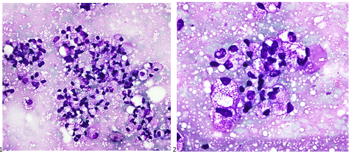
Corynebacterium species are non-motile, facultatively anaerobic, club-shaped gram positive rods that have a characteristic picket fence appearance when stained due to snapping division. These organisms are commonly encountered in clinical samples; however, given their low virulence in general and prevalence as common skin flora they are often considered contaminants or colonizers. The clinical significance of diphtheria toxin (DT)-producing strains has been known for decades. Non-DT-producing  Corynebacterium species were historically considered as non-virulent but in recent years several non-DT-producing Corynebacterium species have been noted to be pathogenic. The wide spread availability of MALDI-TOF MS in most clinical microbiology labs has allowed for rapid and accurate species level identification of Corynebacterium allowing for identification of these pathogenic species in clinical settings. Some Corynebacterium species are highly lipophilic due to the presence of fatty acids, such as mycolic acid, in their cell wall. Isolation of lipophilic Corynebacterium species on standard culture media can be challenging but can be accomplished utilizing blood agar, due to the presence of lipid-containing red cell membranes, with at least 48 hours of incubation. For optimal recovery of lipophilic strains agars supplemented with Tween 80 produce the best recovery rates.

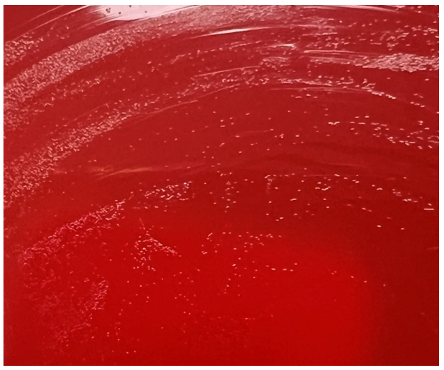
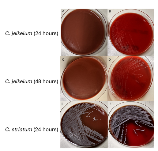
As a lipophilic species, C. jeikeium can be difficult to identify and propagate. It does not grow well on chocolate agar (Fig. 3A & 3C). On blood agar it tends to have scant or dusky growth at 24 hours (Fig. 3B) and appear as translucent, pinpoint colonies at 48 hours (Fig. 3D). Identification using MALDI-TOF can be challenging before 48 hours due to its poor growth on standard media. For comparison, C. striatum, a non-lipophilic Corynebacterium spp. grows relatively well on both blood and chocolate agar. Several Corynebacterium species, including C. jeikeium and C. striatum, are known to be multidrug resistant, especially towards beta-lactam antibiotics.

Figure 3.Images of secondary culture of C. jeikeium and C. striatum inoculated onto chocolate agar (left column) and blood agar (right column) and incubated aerobically at 35°C for 24-48 hours.

Discussion

Corynebacterium jeikeium was initially identified in 1976 and classified by CDC as group JK diptheroids. It is considered part of normal skin flora like most other Corynebacterium spp. In immunosuppressed individuals, especially in persons with hematolymphoid malignancies, invasive disease including dissemination infections have been reported. Infections can also be seen in hardware-associated and prosthetic joint-related infections. Due to its predilection for biofilm formation, C. jeikeium can be difficult to treat in these cases. Cases of infective endocarditis have been reported as well. Like many Corynebacterium species, C. jeikeium is often multidrug resistant. Vancomycin is the first line agent for treatment of infections due to C. jeikeium. Linezolid and daptomycin are other alternative treatment options. When isolating C. jeikeium from blood sources especially in immunocompromised patients like in our case, physicians and other health-care providers must thoroughly assess the total clinical picture before dismissing it as a contaminant.

References

1. Bernard K., Identification of Gram-Positive Bacteria (2023) in AL Leber & CAB Burnham (Eds.) Clinical Microbiology Procedures Handbook (5th ed.) doi:10.1128/9781683670438.CMPH.ch3.18

2. Gupta R, Popli T, Ranchal P, Khosla J, Aronow WS, Frishman WH, El Khoury MY. Corynebacterium Jeikeium Endocarditis: A Review of the Literature. Cardiol Rev. 2021 Sep-Oct 01;29(5):259-262. doi: 10.1097/CRD.0000000000000355. PMID: 32976125.

3. Mattos-Guaraldi AL, Sanchez Dos Santos L, & Vieira VV, Coryneform Gram-Positive Rods (2023) in Carroll et al. (Eds.) Manual of Clinical Microbiology (15th ed.) doi:10.1128/9781683670438.MCM.ch28

4. Moore Pardo SM, Patel RH, Ramsakal A, Greene J. Disseminated Corynebacterium jeikeium Infection in Cancer Patients. Cureus. 2020 Jun 22;12(6):e8764. doi: 10.7759/cureus.8764. PMID: 32714702; PMCID: PMC7377673.

5. Murray BE, Karchmer AW, Moellering RC Jr. Diphtheroid prosthetic valve endocarditis. A study of clinical features and infecting organisms. Am J Med. 1980 Dec;69(6):838-48. doi: 10.1016/s0002-9343(80)80009-x. PMID: 7446550.

6. Ozdemir S, Aydogan O, Koksal Cakirlar F. Biofilm Formation and Antimicrobial Susceptibility of Non-Diphtheria Corynebacterium Strains Isolated from Blood Cultures: First Report from Turkey. Medeni Med J. 2021;36(2):123-129. doi: 10.5222/MMJ.2021.60252. Epub 2021 Jun 18. PMID: 34239764; PMCID: PMC8226407.

7. Tauch A, Kaiser O, Hain T, Goesmann A, Weisshaar B, Albersmeier A, Bekel T, Bischoff N, Brune I, Chakraborty T, Kalinowski J, Meyer F, Rupp O, Schneiker S, Viehoever P, Pühler A. Complete genome sequence and analysis of the multiresistant nosocomial pathogen Corynebacterium jeikeium K411, a lipid-requiring bacterium of the human skin flora. J Bacteriol. 2005 Jul;187(13):4671-82. doi: 10.1128/JB.187.13.4671-4682.2005. PMID: 15968079; PMCID: PMC1151758.

-Arooj Devi MD is a second-year pathology (AP/CP) resident at Brody School of Medicine at East Carolina University. She is interested in breast and gynecological pathology.

-John E. Markantonis DO D(ABMM) is the head of microbiology at ECU Health Medical Center and an assistant professor at Brody School of Medicine at East Carolina University. 

Advances in Determining Time of Death: A Cautionary Note

“Those who cannot remember the past are condemned to repeat it.”

– George Santayana.

Unfortunately, there are many mistakes to learn from if we examine the history of forensic science. Despite being a relatively new discipline, there have been several disastrous failures that were only realized following the advent of DNA analysis – bite mark analysis, tool mark comparison, and arson investigation techniques (to name but a few) have all contributed to past wrongful convictions. Suffice to say that there is historical precedent for “bad” science making its way into the courtroom (see https://innocenceproject.org/misapplication-of-forensic-science/ if you’re interested in reading more).

Lately, I’ve seen several articles about a new method of determining time of death – analysing the “microbial succession” of a decomposing body.1,2 We reviewed the basics of time of death in a previous blog https://labmedicineblog.com/2023/01/25/determining-time-of-death-separating-science-from-pseudoscience/), where we established that estimating postmortem interval is nowhere near as precise as depicted on television. This new technique hopes to change that, and the concept is ingenious. We know the body’s microbiome shifts as postmortem decomposition and putrefaction progress. By measuring and quantifying these changes in different body sites over time (using 16s rRNA sequencing), researchers then identify how our bacterial profiles change. These patterns can then be used to estimate postmortem intervals in cases where it is unknown.

Despite the impressive nature of the preliminary data, I have several reservations about the intent of this research. Many articles discuss microbiome analysis in the context of investigating homicidal deaths, and mentioning this technique in the same sentence as fingerprint and bloodstain evidence draws a direct connection in the readers’ minds to a criminal investigation. It isn’t an unreasonable jump; considering the budgetary limitations of most forensic offices, such an innovative test would likely only be performed in high-stakes cases. If we follow this chain of logic, there is a good probability that this kind of “evidence” would eventually end up as a factor in a homicide trial. When we face the risk of convicting an innocent person, sending them to death row or a life of imprisonment, our excitement around scientific achievement needs to be tempered with pragmatism.  

Research environments are typically well-controlled, in stark contrast to the variety of situations in which people die. This most recent study included 36 cadavers in varying environments; the largest study to date included 63 cadavers had 63.3,4 This sounds like a large number, but imagine the number of variables that need to be considered. Even with attempts to consider factors like soil moisture levels and temperature, the same inevitable problem will arise: each decedent will represent new, unique variables outside of our existing dataset. What if the body has been set on fire? Covered with bleach? Heavily soiled with blood, feces, or vomit? How would gastrointestinal injuries affect the microbiome? Add in varieties of body habitus, baseline commensal bacteria, and environmental variations – the possibilities are nearly endless.

Something can also be statistically significant yet lack practical utility. The reported precision of this method is highly variable between different studies. One recent study estimated time of death within +/- 3 days,3 but other studies have shown higher uncertainty (up to +/- 34 days).4 But how does this error rate compare to our gold standard of investigative context to determine someone’s “window of death”? When were they last seen alive? When did they last text someone, or post on social media? What’s the expiration date on the milk in their fridge? These are methods that seem less “scientific” to a layperson, but they are much more reproducible.

The researchers acknowledge the preliminary nature of these findings, and note further studies are needed. With these admissions, it may sound like my concerns are overly pessimistic. However, even if scientists and pathologists can understand the limitations and nuance, can we also expect lawyers and law enforcement professionals to understand and act accordingly? Most lawyers, judges, and police officers do not have a scientific background.

There may be occasions where a rough estimate is appropriate and helpful to an investigation. If resources eventually allow adoption of microbiome testing on a widespread, affordable basis, I’m sure many families would be interested in knowing what it means for their loved one. But the uncertainty is too high right now to accept microbiome analysis as a tool in criminal proceedings. A high level of scientific scrutiny needs to be applied before any new forensic science techniques are adopted in the courtroom. If this test could possibly be the deciding factor in a person’s innocence or guilt, we need to be absolutely certain the science behind it is quantifiable and reproducible, lest we allow mistakes of the past to be repeated.

REFERENCES:

  1. Barron, Madeline. “Microbial fingerprinting: postmortem microbiome and forensics”. American Society for Microbiology. Published June 3, 2022. Accessed May 18, 2024. https://asm.org/articles/2022/june/microbial-fingerprinting-postmortem-microbiome-and
  1. Schwaiger, Christopher, and LiveScience. “‘Microbiome of death’ uncovered on decomposing corpses could aid forensics”. Scientific American. Published Feb 27, 2024. Accessed May 18, 2024. https://www.scientificamerican.com/article/microbiome-of-death-uncovered-on-decomposing-corpses-could-aid-forensics/#:~:text=’Microbiome%20of%20Death’%20Uncovered%20on%20Decomposing%20Corpses%20Could%20Aid%20Forensics,-Microbes%20that%20lurk&text=The%20same%20%E2%80%9Ckey%20decomposers%E2%80%9D%20show,their%20location%20or%20surrounding%20climate.&text=Microbiology-,Microbes%20that%20lurk%20in%20decomposing%20human%20corpses%20could%20help%20forensic,death%2C%20a%20new%20study%20finds.
  1. Burcham, Z.M., Belk, A.D., McGivern, B.B. et al. A conserved interdomain microbial network underpins cadaver decomposition despite environmental variables. Nat Microbiol 9, 595–613 (2024). https://doi.org/10.1038/s41564-023-01580-y
  1. Tozzo P, Amico I, Delicati A, Toselli F, Caenazzo L. Post-Mortem Interval and Microbiome Analysis through 16S rRNA Analysis: A Systematic Review. Diagnostics (Basel). 2022 Oct 31;12(11):2641. doi: 10.3390/diagnostics12112641. PMID: 36359484; PMCID: PMC9689864.

-Alison Krywanczyk, MD, FASCP, is currently a Deputy Medical Examiner at the Cuyahoga County Medical Examiner’s Office.

It’s Not Always Clear

A 47 year old female presented to the ER with abdominal pain and obstructive jaundice. CT and MRI imaging confirmed a 9.7 cm complex pancreatic head and uncinate process mass that encases and obliterates the distal superior mesenteric vein, encases the superior mesenteric artery, abuts the aorta and IVC and extends into the aortocaval space, displacing the aforementioned structures. By definition, the presentation of this mass indicates locally advanced disease. The patient was referred to gastroenterology for a next-day endoscopy and fine needle aspiration of the pancreatic mass with biliary stent placement.

During the procedure, the cytologist made two smears from two separate passes. The two Diff-Quik-stained smears demonstrated scattered isolated atypical cells with vacuoles in the cytoplasm. The cytologist and pathologist agreed that the cells seen were atypical at best and were debating whether they could be lesional cells or macrophages and that it would be better to see a third smear. The third Diff-Quik smear showed the same cells but this time they were forming aggregates or clusters (Images 1-2). The pathology team deemed the third pass suggestive of tumor. The cytologist collected an additional two needle passes to be rinsed in a balanced salt solution for cell block preparation.

Images 1-2: Pancreas, Head, Endoscopic Ultrasound-guided FNA: DQ-stained smear.

The next morning, the cytologist examined the Papanicolaou-stained slides. The same foamy or microvesicular cytoplasmic vacuoles identified on the Diff-Quik smears were also appreciated on the pap-stained slides (Image 3). There was almost a pink hue to the cytoplasm, which made us question a mucinous tumor. The nuclei were small-to-medium in size with irregular nuclear contours and eccentrically placed. Nucleoli were also appreciated within the tumor cells (Image 4). The H&E-stained cell block sections were beautiful, capturing the nesting architecture of the lesional cells identified on smears (Image 5).

Images 3-5: Pancreas, Head, Endoscopic Ultrasound-guided FNA: 3-4: Pap-stained smear; 5: H&E Cell Block section (400X).

The cytologist and pathologist were both confident that this could be a metastatic renal cell carcinoma, clear cell type. However, there was no evidence of a renal mass. Cancer does what cancer wants though, and to rule out renal cell carcinoma, the pathologist ordered PAX-8 and RCC immunohistochemical (IHC) stains. The tumor cells for both stains came back negative. Back to the drawing board. What if this is an odd presentation of a mucinous cystic neoplasm and these vacuoles are just mucin or a serous cystic neoplasm with glycogen vacuoles? Both mucicarmine and PAS (with and without diastase) were negative, ruling out both neoplasms, respectively. The pathologist and cytologist reviewed the clinical history and imaging. It seems like the only evidence of disease is stemming from the pancreatic mass with moderate lymphadenopathy. At this point, our job is to rule out every primary pancreatic tumor and get this woman an accurate diagnosis so she can 1) get peace of mind, 2) move onto surgery and adjuvant therapy, and 3) finally relieve her excruciating pain.

Let the panel ordering commence! We performed IHC stains on paraffin sections of the cell block with proper positive and negative controls. The tumor cells showed positive staining for AE1/AE3, CK19, CD56, synaptophysin (Image 6), chromogranin (weak), and E-cadherin, membranous staining (but no nuclear staining) for beta-catenin, focal staining for CK7, vimentin, and pCEA, very focal staining (rare cells) for CA19.9, and CD10. In addition to PAX-8 and RCC, the tumor cells showed negative staining for PR and S100. Proliferative index by the Ki-67 immunostain was approximately 2%.

Image 6: Pancreas, Head, Endoscopic Ultrasound-guided FNA: Synaptophysin-positive.

Final diagnosis: Low-grade non-functional pancreatic neuroendocrine tumor (pNET/panNET) with clear cell features.

Had the tumor cells been positive for PR and negative for E-cadherin and chromogranin, a solid pseudopapillary neoplasm may have been in the differential. Negative PAS with diastase also helped to eliminate acinar cell carcinoma from the list of primary pancreatic tumors. It is worth mentioning that the clear cell variant of pNET/panNET is very unusual and may be associated with von Hippel-Lindau disease.

The patient underwent an exploratory laparotomy which revealed metastatic disease. As her symptoms progressively worsened over the next year, the patient decided, using a shared decision-making model with her medical oncologist, to pursue adjuvant therapy. CAPTEM (capecitabine and temozolomide) combined therapy was administered in the setting of progressive metastatic disease. Two years after the initial diagnosis and one years after the initiation of CAPTEM therapy, the patient received 2000 cGy of palliative radiation to the pancreatic mass. While this helped to reduce the disease burden within the primary site, extensive metastasis was noted including an obstructive duodenal mass and multiple liver lesions. Forcep biopsies of the duodenum and both FNAs and core biopsies of the liver demonstrated the same clear cell features of the original tumor, however, the Ki-67 proliferation index increased to 5%, which is consistent with an intermediate or grade 2 (G2) pNET. The patient developed uncontrolled bleeding and subsequently severe anemia and went on hospice. Within a matter of weeks after stabilizing her hemoglobin, the patient came off of hospice and began treatment with Lutathera, a radioligand therapy or peptide receptor radionuclide therapy (PRRT) which targets cells with somatostatin receptors. This therapy kept her relatively stable for an additional two years, but imaging demonstrated further progression on PRRT so an mTOR inhibitor was introduced to reduce the disease burden.

The beauty of oncology is that new therapies and advancements are always being developed and released, often improving the patient’s longevity and/or quality of life. The beauty of pathology is that the diagnosis is not always clear, but using the ever-expanding diagnostic and prognostic tools available to us can help us give the patient an answer and guide future treatments.

-Taryn Waraksa-Deutsch, MS, SCT(ASCP)CM, CT(IAC), has worked as a cytotechnologist at Fox Chase Cancer Center, in Philadelphia, Pennsylvania, since earning her master’s degree from Thomas Jefferson University in 2014. She is an ASCP board-certified Specialist in Cytotechnology with an additional certification by the International Academy of Cytology (IAC). She is also a 2020 ASCP 40 Under Forty Honoree.

Microbiology Case Study: A Friend or Foe with No Cell Wall

A 28 year old woman underwent an elective myomectomy for menorrhagia caused by fibroids. Postoperatively, she developed fevers. A CT scan of the abdomen and pelvis showed a complex pelvic fluid collection measuring 5.4 by 4.4 by 7.0 cm. Drainage was attempted by Interventional Radiology without success. She then underwent an exploratory laparotomy with drainage of the collection, evacuation of a hematoma, and removal of an IUD. The abscess fluid was sent to the Microbiology lab for culture.

The Gram stain of the fluid revealed 1+PMN with no organisms. After 48 hours of incubation, there was few to moderate growth of pinpoint clear colonies on blood agar, with characteristic miniscule appearance of a central area of dense growth and peripherally less dense (Figure 1). Upon closeup reviewing of the colonies revealed a “fried egg” appearance (Fig 2).

There was no growth on MacConkey plate. Final identification by MALDI-TOF demonstrated Mycoplasma hominis.

Figure 1. Blood agar plate growing clear small colonies of M. hominis.
Figure 2. Characteristic colonies of organism with peripheral growth and denser central growth with fried-egg appearance.

Discussion

Mycoplasma hominis is a facultative anaerobe of the Mycoplasma genus, which is among the smallest free-living organisms known. They are fastidious and differentiated from other bacteria by their small size and absence of cell walls. Infections with M. hominis predominately involve the urogenital tract of females, causing pelvic inflammatory disease, bacterial vaginosis, and postpartum fever.1 Extragenital manifestations of M. hominis infections are rare but include extragenital abscesses, CNS infections in neonates, and septic arthritis.2 Reports of Mycoplasma hominis infections vary based on demographics (country, age, and number of sexual partners) but have been isolated in 4 to 30% of urogenital infections in females. 1,3

The lack of cell walls confers both diagnostic and clinical challenges. Mycoplasmas is not seen on the routine Gram stain smears. Instead of a cell wall, they have a triple-layered membrane composed of sterol.3 When able to grow in culture, colonies are small and have a ‘fried egg’ appearance on agar.4 Of the Mycoplasma species, M. hominis is the least fastidious and the most common Mycoplasma isolated on conventional culture agar media. 4-6 Non-traditional culture-based diagnosis is often made via molecular testing. 7 It is often detected in coinfections with Trichomonas vaginalis and thought to be a symbiotic relationship. 8-9

Since M. hominis is also found to be colonized in the genital tract of normal healthy individuals, it can be challenging to establish the clinical and diagnostic significance of M. hominis. With evolving molecular diagnostic assays targeting STI agents, it has been a controversial topic for assay manufacturers or labs that develop their own in-house assays whether there is a clinical utility for M. hominis PCR as part of STI multiplex PCR panels.

In contrast to Mycoplasma pneumoniae, M. hominis has intrinsic resistance to macrolides. 10 Preferred treatment regimens for M hominis infections include tetracyclines, clindamycin and fluoroquinolones. 11 However, resistance to members of these antibiotic classes has been reported and differs based on country of origin. 11 The absence of a cell wall explains the inherent resistance of Mycoplasma species to the beta-lactam antibiotic class. Its isolation in coinfections, particularly Trichomonas has been controversial in its contribution to emerging Trichomonas metronidazole resistance. 12

References

  1. Verteramo, R., Pastella, A., Calzolari, E., et al. An epidemiological survey of Mycoplasma hominis and Ureaplasma urealyiticum in gynaecological outpatients, Rome, Italy. Epidemiology & Infection,2013, 141(12), 2650-2657
  2. Zheng X, Olson DA, Tully JG, Watson HL, Cassell Gh, Gustafson DR, Svien KA, Smith TF: Isolation of Mycoplasma hominis from a brain abscess. J Clin Microbiol. 1997, 35: 992-994.
  3. Thomas Prescott Atkinson, Mitchell F. Balish, Ken B. Waites, Epidemiology, clinical manifestations, pathogenesis and laboratory detection of Mycoplasma pneumoniae infections, FEMS Microbiology Reviews, Volume 32, Issue 6, November 2008, Pages 956–973
  4. Razin S. Mycoplasmas. In: Baron S, editor. Medical Microbiology. 4th edition. Galveston (TX): University of Texas Medical Branch at Galveston; 1996. Chapter 37.
  5. Meloni GA, Bertoloni G, Busolo F, Conventi L. Colony morphology, ultrastructure and morphogenesis in Mycoplasma hominis, Acholeplasma laidlawii and Ureaplasma urealyticum. J Gen Microbiol. 1980;116(2):435-443.
  6. Christiansen G, Jensen LT, Boesen T, Emmersen J, Ladefoged SA, Schiotz LK, Birkelund S: Molecular biology of Mycoplasma. Wien Klin Wochenschr. 1997, 109: 557-561
  7. Baczynska, A., Svenstrup, H.F., Fedder, J. et al. Development of real-time PCR for detection of Mycoplasma hominisBMC Microbiol 4, 35 (2004).
  8. Tine RC, Dia L, Sylla K, Sow D, Lelo S, Ndour CT. Trichomonas vaginalis and Mycoplasma infections among women with vaginal discharge at Fann teaching hospital in Senegal. Trop Parasitol. 2019 Jan-Jun;9(1):45-53.
  9. Vancini, R.G., Benchimol, M. Entry and intracellular location of Mycoplasma hominis in Trichomonas vaginalis . Arch Microbiol 189, 7–18 (2008).
  10. Pereyre S, Gonzalez P, De Barbeyrac B, Darnige A, Renaudin H, Charron A, Raherison S, Bébéar C, Bébéar CM. Mutations in 23S rRNA account for intrinsic resistance to macrolides in Mycoplasma hominis and Mycoplasma fermentans and for acquired resistance to macrolides in M. hominis. Antimicrob Agents Chemother. 2002 Oct;46(10):3142-50.
  11. Krausse R, Schubert S. In-vitro activities of tetracyclines, macrolides, fluoroquinolones and clindamycin against Mycoplasma hominis and Ureaplasma ssp. isolated in Germany over 20 years. Clin Microbiol Infect. 2010;16(11):1649-1655.
  12. Dessì, D., Margarita, V., Cocco, A., Marongiu, A., Fiori, P., & Rappelli, P. (2019). Trichomonas vaginalis and Mycoplasma hominis: New tales of two old friends. Parasitology, 146(9), 1150-1155. doi:10.1017/S0031182018002135

-Dr. Alex Shaffer is a first-year ID fellow (2023-2025) at Montefiore Einstein. Alex is interested in the diagnostic stewardship projects and has been actively involved in various activities with ID and micro lab faculty.

-Phyu Thwe, Ph.D, D(ABMM), MLS(ASCP)CM is Associate Director of Infectious Disease Testing Laboratory at Montefiore Medical Center, Bronx, NY. She completed her medical and public health microbiology fellowship in University of Texas Medical Branch (UTMB), Galveston, TX. Her interests includes appropriate test utilization, diagnostic stewardship, development of molecular infectious disease testing, and extrapulmonary tuberculosis.