Microbiology Case Study: A 46 Year Old with Chest Pain

Case History

A 46 year old male with a history of cystic fibrosis and bilateral lung transplant two years prior presented to the hospital with chest pain and hemoptysis. The patient was recently diagnosed with COVID-19, and a CT chest revealed multiple rounded, mass-like opacities with central cavitation. As imaging was not consistent with COVID-19 pulmonary disease and no clear risk for tuberculosis could be identified, a bronchoscopy with transbronchial biopsy was performed. Tissue and bronchiolar lavage fluid were collected and submitted to the microbiology laboratory for analysis. Viral etiologies including influenza A/B, Parainfluenza 1-3, Adenovirus, RSV and metapneumovirus were ruled out through molecular studies. Galactomannan was negative from the BAL fluid, as were fungal and mycobacterial cultures and Mycobacterium tuberculosis PCR. GMS staining of the biopsy was negative but organizing pneumonia and mononuclear infiltrate was noted. The patient had a history of recurrent multidrug-resistant Pseudomonas aeruginosa infection and was being managed with empiric ceftazidime/avibactam.

Laboratory Identification

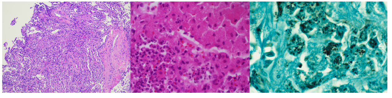
Gram stains of both the tissue and BAL fluid were generally unremarkable. Histopathological analysis of the transbronchial tissue revealed changes suggestive of organizing pneumonia with mononuclear infiltrate (Image 2, left). Bacterial growth of a predominant organism from both the BAL and biopsy tissue was observed on plates after 48 hours on blood and chocolate agars but was absent on MacConkey agar. At 96 hours, the colonies of the organism had become mucoid, slightly pink and had coalesced (Image 1, right). Gram staining of the growth revealed short, poorly staining gram positive coccobacilli with a beaded appearance. Due to the incomplete gram staining of this isolate, modified acid-fast staining was attempted which was positive (Image 1, left). The organism was both catalase- and urease-positive. The isolate was subsequently identified by MALDI-TOF MS as Rhodococcus equi, and the patient was discharged from the hospital on imipenem and linezolid.

Image 1. (Left) Modified acid-fast (MAF) staining revealing small, MAF-positive coccobacilli (black arrowheads).  (Right) Characteristic, mucoid salmon-colored colonies of the isolate on blood agar after 96 hours incubation. ​
Image 2. (Left) Transbronchial biopsy revealing areas of histiocyte aggregation and mononuclear infiltrate (H&E, 10X magnification).  (Middle) Representative image of expanded histiocytes with small, pale-staining round forms in a background of neutrophils (H&E, 40X magnification).  (Right) Representative image of histiocytes filled with coccoid and coccobacilliary forms (GMS, 40X magnification).​

Discussion

Rhodococcus equi is a zoonotic pathogen which primarily causes infections among immunocompromised hosts. Infrequently isolated clinically, the organism is a primary pathogen of horses causing pneumonia with abscess formation in foals, often with dissemination into peripheral sites due to high organism burden. The organism is excreted in feces of infected animals, leading to contamination of soils from farms, ranches, and other agricultural environments from which the organism is either aerosolized and inhaled or acquired via direct inoculation.1 While human infections are classically associated with exposure to horses or their environment, there is a growing body of literature to suggest that many patients with microbiologically proven cases of R. equi infection lack such environmental exposures. This patient falls into the latter category, with no known exposure to livestock.

                R. equi is a member of the aerobic actinomycetes. Like Nocardia sp., the cell wall of R. equi contains mycolic acids which lead to positivity when stained with a modified acid-fast stain. The organism is a facultative, intracellular pathogen surviving within macrophages and histiocytes, leading to granulomatous inflammation, eventually leading to necrosis.2 Immunosuppression (including HIV infection or immunosuppressive therapy) is a major risk factor for R. equi infection, as most clinical cases are reported in this setting. In immunocompromised hosts, the spectrum of disease manifestations of R. equi are diverse, but most commonly (approx. 80%) include pulmonary involvement3 with upper lobe cavitary pneumonia.4 Characteristic malakoplakia (an infiltration of foamy histocytes with intracellular bacteria and basophilic inclusions name Michaelis-Gutmann bodies)1 can be associated with R. equi infection. These structures were noticeably absent in this patient’s case despite the observed histocyte aggregation and mononuclear infiltrate (Image 2, center, left).

R. equi pneumonia among solid organ transplant recipients, such as the patient in this case is associated with low overall morbidity and mortality, but require protracted antibiotic therapy regimens.1 Susceptibility testing is warranted to guide therapy of R. equi due to unpredictable resistance patterns among isolates. This patient’s isolate was revealed to be susceptible to amoxicillin/clavulanate, ceftriaxone, imipenem, ciprofloxacin, moxifloxacin, clarithromycin, amikacin, tobramycin, minocycline, trimethoprim/sulfamethoxazole, vancomycin, linezolid, and rifampin. The patient was discharged on imipenem/linezolid. At follow-up, the patient had clinically improved with a resolution of symptoms, but his radiologic abnormalities persisted and thus remains on oral therapy with moxifloxacin and minocycline.

References

Yamshchikov, AV, Schuetz, A, and Lyon, GM. Rhodococcus equi infection. 2010. Lancet Infect. Dis. 10:350-359.

Prescott, JF. Rhodococcus equi: an Animal and Human Pathogen. 1991. Clin. Microbiol. Rev. 4(1):20-34.

Weinstock, DM, and Brown, AE. Rhodococcus equi: an emerging pathogen. 2002. Clin. Infect. Dis. 34:1379-1385.

Mutaner, L, et. al. Radiologic featuresof Rhodococcus equi pneumonia in AIDS. Eur. J. Radiology. 1997. 66-70.

-Andrew Clark, PhD, D(ABMM) is an Assistant Professor at UT Southwestern Medical Center in the Department of Pathology, and Associate Director of the Clements University Hospital microbiology laboratory. He completed a CPEP-accredited postdoctoral fellowship in Medical and Public Health Microbiology at National Institutes of Health, and is interested in antimicrobial susceptibility and anaerobe pathophysiology.

-Dominick Cavuoti is a Professor in the Department of Pathology at UT Southwestern Medical Center. Dr. Cavuoti is a board certified AP/CP who is a practicing Clinical Microbiologist, Infectious Disease pathologist and Cytopathologist.


-Clare McCormick-Baw, MD, PhD is an Assistant Professor of Clinical Microbiology at UT Southwestern in Dallas, Texas. She has a passion for teaching about laboratory medicine in general and the best uses of the microbiology lab in particular.

Microbiology Case Study: A 17 Year Old with Chest Pain

Case History

A 17 year old female who presented to the emergency department with complaints of fever, vomiting, diarrhea, and chest pain for the past two weeks. She also reported an unintentional weight loss of 20 lbs. Her medical history consisted of essential hypertension for which she was previously on medication, however had been discontinued two years ago due to normal blood pressure. The patient reported that she is sexually active with one male partner and denied use of protection. She denied any other sexual partners or any prior history of sexually transmitted infections. Her urine NAAT testing was positive for chlamydia, but negative for gonorrhea. Blood cultures collected at the time of admission resulted in growth of gram-negative diplococci on day 2 of admission (Image 1) and colony growth on chocolate agar (Image 2). The organism was positive for both catalase and oxidase and identified by matrix-assisted light desorption ionization- time of flight (MALDI-TOF) as Neisseria gonorrhoeae. Due to her chest pain complaints and QT prolongation on EKG, a trans-thoracic echo was performed that demonstrated a large aortic root abscess suggestive of infective endocarditis. Ceftriaxone was started as treatment for her gram-negative endocarditis, and she was emergently transferred to another facility where an aortic valve replacement and patch aortoplasty were performed.

Image 1. Gram stain of the blood culture showing gram negative diplococci.
Image 2. Neisseria gonorrhoeae on chocolate agar producing small gray-white colonies.

Discussion

Neisseria gonorrhoeae is a fastidious, oxidase positive, gram negative diplococcus, commonly transmitted through sexual contact.2,3 Neisseria uniquely grows on chocolate agar and VPN/Thayer Martin agar, and has virulence factors such as pilli that attach to mucosal surfaces, and many antigenic variations that make it a highly resistant organism prone to reinfection.

In the laboratory, N. gonorrhea grows well on chocolate agar after 24-48 hours of incubation (Image 2) with less robust or no growth on blood agar. It is positive for both catalase and oxidase. Traditionally, sugar fermentation was used to differentiate Neisseria species from one another, but more ore rapid identification methods (MALDI-TOF and PCR) are being increasingly used in most clinical laboratories

In men, Neisseria usually ascends the genitourinary tract to cause prostatitis. In women, the infection can disseminate to cause pelvic inflammatory disease, which can cause scarring in the fallopian tubes, resulting in infertility. Neisseria also can present as an asymmetric polyarthritis, most commonly to the knees. The main treatment of Neisseria gonorrhea is ceftriaxone. Gentamicin is an acceptable alternative in patients with severe cephalosporins allergy.

This case involves a rare presentation of infective endocarditis caused by disseminated gonorrhea infection. Previous reported cases of gonococcal endocarditis1,4 reported ad subacute presentation in around 2-4 weeks with generalized fatigue, fevers, arthritis, rash, renal dysfunction, and new cardiac murmurs. Because it can present without preceding genitourinary symptoms, disseminated gonorrheal can be difficult to recognize. The infection is usually aggressive, forming large vegetations and rapid valve destruction, despite antibiotic treatment. Most commonly it involves the aortic valve, as seen in the case presented above, but can also involve the mitral and tricuspid valves in some cases. The damage usually requires valve replacement surgery in addition to antimicrobial therapy.5,6 Lastly, this case demonstrates the limitations of the urine NAAT to diagnose gonorrhea specifically in females and/or asymptomatic patients due the possible presence of inhibitors and the need for further testing if clinical suspicion remains.7

References

  1. Said M, Tirthani E. Gonococcal Infective Endocarditis Returns. Cureus. 2021 Sep 14;13(9):e17955. doi: 10.7759/cureus.17955. PMID: 34660143; PMCID: PMC8515499.
  2. Ryan, K. J., Ray, G., and Sherris, J. C. (2004). Sherris Medical Microbiology: An introduction to Infectious Diseases, 4th edition. McGraw-Hill Medical.
  3. Centers for Disease Control and Prevention. Gonorrhea. Available from: https://www.cdc.gov/std/gonorrhea/stdfact-gonorrhea-detailed.htm. Last updated 2021 July 22; cited on 2022 March 21.
  4. Fenech, Marylou, et al. “Neisseria Gonorrhoeae Infective Endocarditis.” BMJ Case Reports, BMJ Specialist Journals, 1 May 2022
  5. Thompson EC, Brantley D. Gonoccocal endocarditis. J Natl Med Assoc. 1996 Jun;88(6):353-6. PMID: 8691495; PMCID: PMC2608094.
  6. Nie S, Wu Y, Huang L, Pincus D, Tang YW, Lu X. Gonococcal endocarditis: a case report and literature review. Eur J Clin Microbiol Infect Dis. 2014 Jan;33(1):23-7. doi: 10.1007/s10096-013-1921-x. Epub 2013 Jul 16. PMID: 23856883.
  7. Whiley DM, Tapsall JW, Sloots TP. Nucleic acid amplification testing for Neisseria gonorrhoeae: an ongoing challenge. J Mol Diagn. 2006 Feb;8(1):3-15. doi: 10.2353/jmoldx.2006.050045. PMID: 16436629; PMCID: PMC1871692.

-Olivia Piscano is a second-year medical student at the Medical College of Georgia. She is currently interested in Internal Medicine, Pediatrics, and Infectious Disease.

-Hasan Samra, MD, is the Director of Clinical Microbiology at Augusta University and an Assistant Professor at the Medical College of Georgia.

Microbiology Case Study: 57 year old Female with Altered Mental Status and Declining Health

Case Description

A 57 year old female presents to the emergency department with altered mental status, decreased appetite and chest, abdominal and pelvic pain. She has a complex medical history including end-stage renal disease, cardiovascular disease with pacemaker placement, and recurrent ascites. Her physical exam was notable for hypotension (65/52), hypothermia (31.7°C), and abdominal distention. A CT of her abdomen and pelvis revealed marked ascites, and bloodwork indicated leukocytosis (12.81, ref 4.22-10.33), elevated lactate (4.1, ref 0.5-2.2), and acidemia (7.27). Given the concern for septic shock, an infectious workup was initiated. A diagnostic paracentesis was undertaken which revealed rare yeast forms by Gram stain (Figure 1). Routine and fungal cultures of the ascites fluid grew yeast which was identified as Cryptococcus neoformans by matrix-assisted laser desorption/ionization-time-of-flight mass spectrometry (MALDI-TOF MS). Bacterial blood cultures turned positive after 3 days and Cryptococcus neoformans was identified (Figure 2). Interestingly, serum cryptococcal antigen tests were negative.

The patient was treated with amphotericin B, vancomycin, and meropenem. Despite intervention, the patient’s clinical condition continued to deteriorate, leading to multiple organ failure. The patient was transitioned to comfort care and expired soon thereafter.

Figure 1. Gram stain of peritoneal fluid (ascites) demonstrating two yeasts with surrounding capsule and host cells (100x objective, oil immersion).
Figure 2.  Bacterial blood culture with encapsulated yeasts on Gram stain (100x objective, oil immersion).

Discussion

This case highlights two significant points: 1) atypical presentation of a cryptococcal infection and 2) the value of complimentary approaches in the diagnostic workup of an infectious etiology.

Cryptococciare environmental fungi with worldwide distribution, classily causing opportunistic central nervous system infection in patients with either uncontrolled HIV or other significantly immunocompromising conditions. In this patient population, a staggering 70-90% of cryptococcal infections manifest as meningitis, with a one-year mortality rate as high as 70% in some regions.1 Meningitis caused by Cryptococcus sp. accounts for nearly 1 in 7 HIV-related deaths worldwide.2 This case and others,3 serve as an important reminder that cryptococcal disease may present in different ways, including peritonitis in a patient without significant immunosuppression.

Several methods are available to aid in the identification of Cryptococcus sp.4 Direct microscopic evaluation of Cryptococcus in fluid and tissue samples reveals round, narrow-based budding yeast of variable size (2-20 um). The capsule of Cryptococcus is classically seen with India ink, though recent work has shown that Gram stain is as effective.5 In our patient, direct microscopy of the peritoneal fluid provided the first clue that Cryptococcus was the causative agent. Other useful stains used for histopathological analysis include mucicarmine and Fontana-Masson, which stain the capsule and melanin, respectively. In culture, strains of Cryptococcus sp. typically elaborate a robust capsule leading to the formation of mucoid colonies. However, acapsular strains have also been identified.

Biochemical hallmarks of Cryptococcus sp. include the production of urease and phenoloxidase leading to the formation of melanin which is absorbed into the cell wall. Phenoloxidase activity is exploited for diagnostic purposes as it leads to melanized pigmentation in the presence of caffeic acid, such as in either a caffeic acid disk test or on Bird Seed agar.

Non-culture-based methods for the diagnosis of cryptococcal infections include detection of cryptococcal capsular antigens by either ELISA, latex agglutination, or a lateral flow immunoassay (LFA). The LFA is a cost-effective test with rapid turnaround time which exhibits strong agreement with other antigen detection methods. While the LFA has proven useful in a variety of settings,6 our patient’s LFA was negative, underlining the importance of using orthogonal methods in parallel to identify microbes. In cases where patients are infected with an acapsular strain of Cryptococcus, the antigen testing will be negative since the antigenic target (i.e the capsule) is missing. An important consideration when assessing discrepancies between Gram stain/culture and ancillary immunoassay testing is “prozone effect”. Prozone, or Hook effect, is a phenomenon where overwhelming amounts of analyte impairs immunocomplex formation, causing a lack of analyte detection (i.e false-negative test). To account for prozone, a serial dilution series of the sample with repeat testing should be performed to ensure accurate correlation.

References

  1. World Health Organization. Guidelines for the diagnosis, prevention, and management of cryptococcal disease in HIV-infected adults, adolescents and children, March 2018: supplement to the 2016 consolidated guidelines of the use of antiretroviral drugs for treating and preventing HIV infection.
  2. Rajasingham R, Smith RM, Park BJ, Jarvis JN, Govender NP, Chiller TM, Denning DW, Loyse A, Boulware DR. Global burden of disease of HIV-associated cryptococcal meningitis: an updated analysis. The Lancet infectious diseases. 2017 Aug 1;17(8):873-81.
  3. El-Kersh K, Rawasia WF, Chaddha U, Guardiola J. Rarity revisited: cryptococcal peritonitis. Case Reports. 2013 Jul 10;2013:bcr2013009099.
  4. Mais DD. Quick compendium of clinical pathology. American Society for Clinical Pathology Press; 2018.
  5. Coovadia YM, Mahomed S, Dorasamy A, Chang C. A comparative evaluation of the Gram stain and India ink stain for the rapid diagnosis of cryptococcal meningitis in HIV infected patients in Durban: brief report. Southern African Journal of Infectious Diseases. 2015 Jan 1;30(2):61-3.
  6. Perfect JR, Bicanic T. Cryptococcosis diagnosis and treatment: What do we know now. Fungal Genetics and Biology. 2015 May 1;78:49-54.

AUTHORS

-Andrew T. Nelson, MD, PhD, is a Clinical Pathology resident at UT Southwestern Medical Center in his second year. He has an interest in Clinical Chemistry.

-Andrew Clark, PhD, D(ABMM) is an Assistant Professor at UT Southwestern Medical Center in the Department of Pathology, and Associate Director of the Clements University Hospital microbiology laboratory. He completed a CPEP-accredited postdoctoral fellowship in Medical and Public Health Microbiology at National Institutes of Health, and is interested in antimicrobial susceptibility and anaerobe pathophysiology.

-Clare McCormick-Baw, MD, PhD is an Assistant Professor of Clinical Microbiology at UT Southwestern in Dallas, Texas. She has a passion for teaching about laboratory medicine in general and the best uses of the microbiology lab in particular.

An Adult Patient Presents with Mild Penile Irritation and Discharge

Patient History

An adult male presented to the primary care office with mild penile irritation and discharge without fever, dysuria, or other lesions. He is sexually active and reported recent unprotected sex with multiple partners. He is on pre-exposure prophylaxis for HIV and tested non-reactive for HIV, HCV, and syphilis antibodies. Chlamydia and gonorrhea were detected in his urine, rectal, and throat specimens by PCR. The lab paged the director to review and verify the results. Is it possible to be positive for both chlamydia and gonorrhea?

Discussion

In the United States, chlamydia and gonorrhea are the most commonly reported sexually transmitted bacterial infections. While most cases of chlamydia and gonorrhea are sexually transmitted, neonates can become infected by perinatal transmission.1,2,3 To prevent long-term complications in women, all sexually active women aged <25 years and older women with increased risk of infection should get tested annually for chlamydia and gonorrhea. All pregnant women <25 years old or are considered high risk should be screened at the first prenatal visit and in the third trimester or at the time of delivery for both organisms. CDC recommends screening genital and extragenital sites at least annually for all sexually active MSM at risk for infection.4

Chlamydia trachomatis (C. trachomatis) is a gramvnegative, obligate, aerobic, coccoid or rod shape bacteria that does not grow in routine culture. C. trachomatis cannot synthesize ATP and humans are the only known natural host for C. trachomatis.4 Neisseria gonorrhoeae (N. gonorrhoeae) is a Gram-negative, facultatively intracellular, obligate aerobe diplococci. While this organism can be grown in culture, sensitivity is lower compared to routine molecular methods. Co-infection is common, with an estimated 10–40% of patients with gonorrhea also infected with chlamydia, and the data also suggested an interplay between these two pathogens. 5,6,7 Patients with chlamydia and gonorrhea co-infection can have increased gonococcal bacterial load, which might facilitate gonorrhea transmission compared with a single infection. Chlamydia can evade the host immune response by preventing neutrophil extracellular traps (NETs) production, which can help gonorrhea to establish intracellular infection.8 Studies in mice suggest that C. trachomatis induces changes in the genital tract immune environment, making it a more permissive environment for N. gonorrhoeae.9

Appropriate specimens include self- or clinician-collected vaginal swab, endocervical swab, urethral swab, and first catch urine. For chlamydial and gonococcal infection diagnosis, CDC recommends testing by nucleic acid amplification tests (NAATs). NAATs are more sensitive and specific compared to other methods. FDA has approved NAATs for urogenital specimens and only particular platforms are approved for rectal and oropharyngeal specimens. C. trachomatis does not grow in routine culture and diagnosis at this time relies solely on NAAT. For N. gonorrhoeae, culture and antibiotic susceptibility should be evaluated in case of suspected treatment failure. Our lab uses Abbott Real-time CT/NG assay, which is currently FDA approved for testing urogenital specimens only.

CDC recommends treating chlamydia with a seven-day course of doxycycline with sexual abstinence until treatment completion/resolution of symptoms. Azithromycin or levofloxacin can be used as alternatives. For gonorrhea, a single ceftriaxone intramuscular injection is recommended, and gentamicin with azithromycin can be used in case of cephalosporin allergy. Unfortunately, for pharyngeal gonorrhea, there is no reliable alternative available for ceftriaxone allergy. Sexual partner evaluation, testing, and presumptive treatment are recommended, along with patient treatment.10 In cases where the chlamydial infection has not been ruled out, patients should also receive anti-chlamydial therapy. A test-of-cure (follow-up testing) for gonorrhea is required in throat infections only after 14 days of the treatment.10

References:

  1. Kreisel KM, Spicknall IH, Gargano JW, Lewis FM, Lewis RM, Markowitz LE, Roberts H, Satcher Johnson A, Song R, St. Cyr SB, Weston EJ, Torrone EA, Weinstock HS. Sexually transmitted infections among US women and men: Prevalence and incidence estimates, 2018. Sex Transm Dis 2021; in press.
  2. CDC. Sexually Transmitted Disease Surveillance, 2020. Atlanta, GA: Department of Health and Human Services; April 2022.
  3. https://www.cdc.gov/std/chlamydia/stdfact-chlamydia-detailed.
  4. https://www.cdc.gov/std/treatment-guidelines/msm.
  5. Creighton S, Tenant-Flowers M, Taylor CB, Miller R, Low N. Coinfection with gonorrhea and chlamydia: how much is there and what does it mean? Int J STD AIDS. 2003; 14:109–13.
  6. Althaus CL, Turner KM, Mercer CH, Auguste P, Roberts TE, Bell G, Herzog SA, Cassell JA, Edmunds WJ, White PJ, Ward H, Low N. Effectiveness and cost-effectiveness of traditional and new partner notification technologies for curable sexually transmitted infections: observational study, systematic reviews and mathematical modelling. Health Technol Assess. 2014 Jan;18(2):1-100, vii-viii. doi: 10.3310/hta18020. PMID: 24411488; PMCID: PMC4780998.
  7. Creighton S. Gonorrhoea. BMJ Clin Evid. 2014:2014.
  8. Rajeeve K, Das S, Prusty BK, Rudel T. Chlamydia trachomatis paralyses neutrophils to evade the host innate immune response. Nat Microbiol. 2018 Jul;3(7):824-835. doi: 10.1038/s41564-018-0182-y. Epub 2018 Jun 25. PMID: 29946164.
  9. Vonck RA, Darville T, O’Connell CM, Jerse AE. Chlamydial infection increases gonococcal colonization in a novel murine coinfection model. Infect Immun. 2011 Apr;79(4):1566-77. doi: 10.1128/IAI.01155-10. Epub 2011 Jan 18. PMID: 21245268; PMCID: PMC3067530.
  10. St. Cyr S, Barbee L, Workowski KA, et al. Update to CDC’s Treatment Guidelines for Gonococcal Infection, 2020. MMWR Morb Mortal Wkly Rep 2020;69:1911–1916. DOI: http://dx.doi.org/10.15585/mmwr.mm6950a6external icon

-Payu Raval, MD is a 2nd year anatomic and clinical pathology resident at University of Chicago (NorthShore). Her academic interests include hematology, molecular, and surgical pathology.

-Paige M.K. Larkin, PhD, D(ABMM), M(ASCP)CM is the Director of Molecular Microbiology and Associate Director of Clinical Microbiology at NorthShore University HealthSystem in Evanston, IL. Her interests include mycology, mycobacteriology, point-of-care testing, and molecular diagnostics, especially next generation sequencing.